. 2012 Jul;18(7):1060-8.
doi: 10.1038/nm.2811.

Hepatitis B virus-induced lipid alterations contribute to natural killer T cell-dependent protective immunity

Affiliations

Hepatitis B virus-induced lipid alterations contribute to natural killer T cell-dependent protective immunity

Sebastian Zeissig et al. Nat Med. 2012 Jul.

Abstract

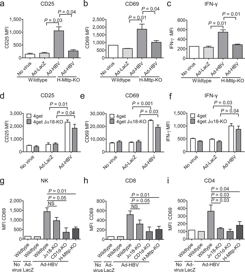
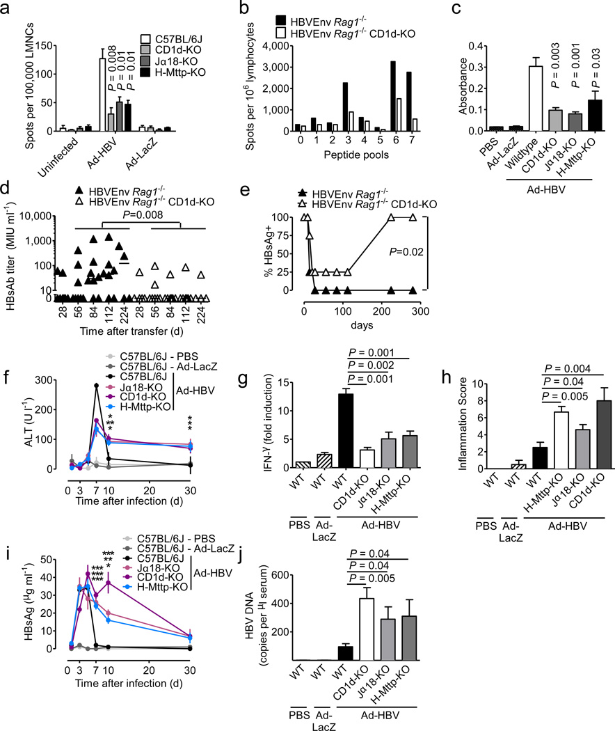
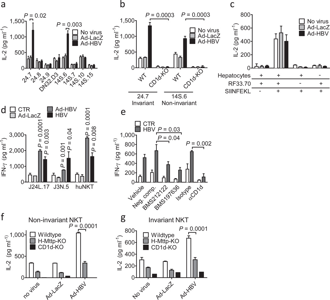
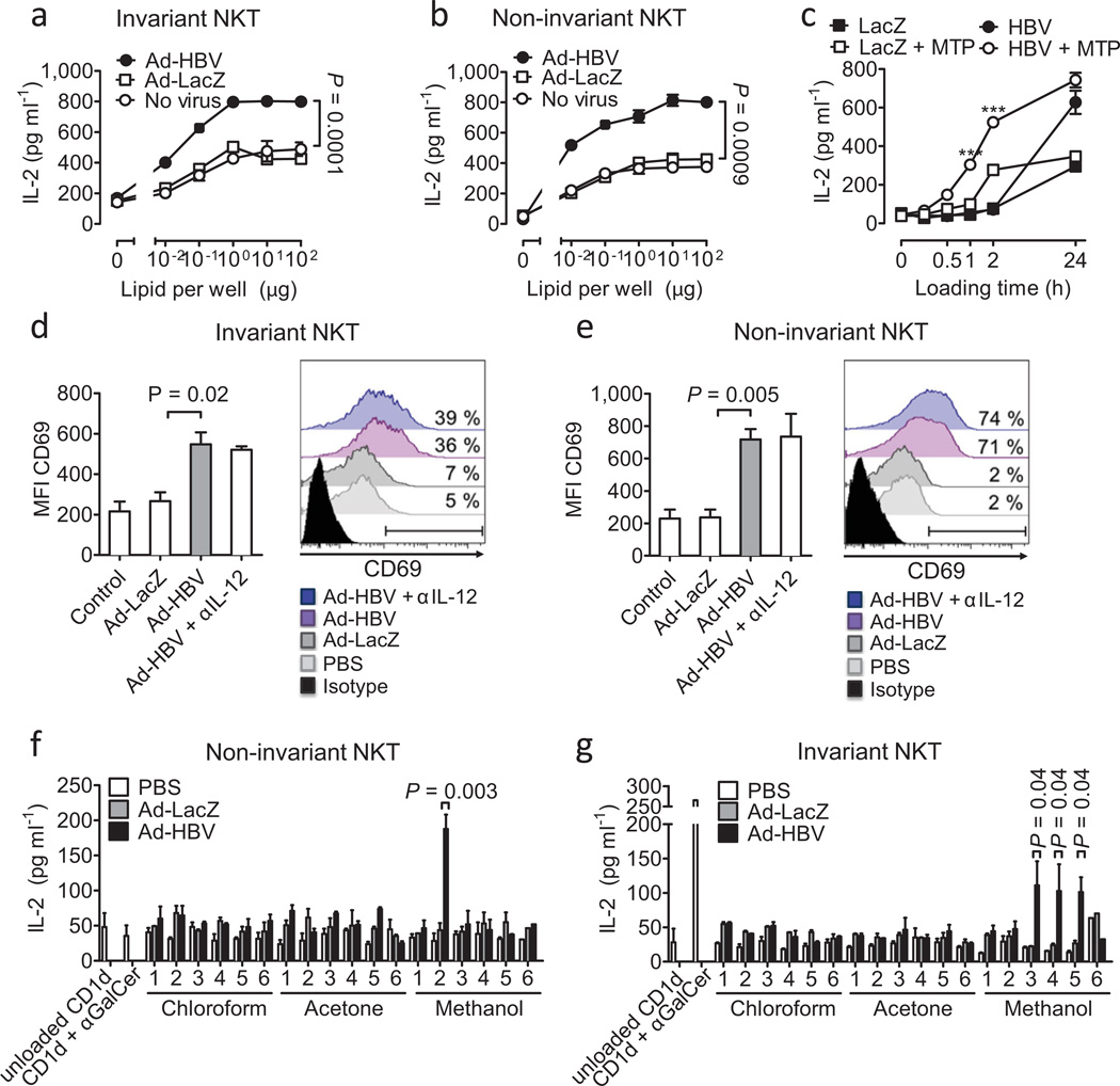
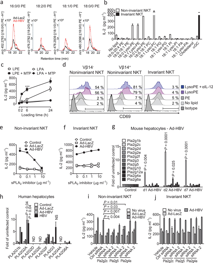
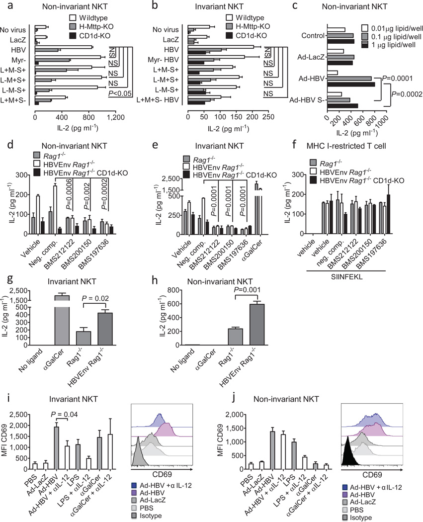
In most adult humans, hepatitis B is a self-limiting disease leading to life-long protective immunity, which is the consequence of a robust adaptive immune response occurring weeks after hepatitis B virus (HBV) infection. Notably, HBV-specific T cells can be detected shortly after infection, but the mechanisms underlying this early immune priming and its consequences for subsequent control of viral replication are poorly understood. Using primary human and mouse hepatocytes and mouse models of transgenic and adenoviral HBV expression, we show that HBV-expressing hepatocytes produce endoplasmic reticulum (ER)-associated endogenous antigenic lipids including lysophospholipids that are generated by HBV-induced secretory phospholipases and that lead to activation of natural killer T (NKT) cells. The absence of NKT cells or CD1d or a defect in ER-associated transfer of lipids onto CD1d results in diminished HBV-specific T and B cell responses and delayed viral control in mice. NKT cells may therefore contribute to control of HBV infection through sensing of HBV-induced modified self-lipids.


Figures

Figure 1
Figure 1
NKT cells become activated in response to Ad-HBV and contribute to adaptive immune responses. Expression of the indicated markers on hepatic iNKT cells (a–c, αGalCer/CD1d+CD3+) and non-invariant NKT cells (d–f, 4Get X Jα18-KO) two days after i.v. administration of PBS (no virus), Ad-HBV or Ad-LacZ as determined by flow cytometry. In a–c, H-Mttp-KO and wildtype mice are shown. (g–i) NK (CD3 NK1.1+) and conventional (αGalCer/CD1d) CD8+ and CD4+ T cell activation five days after virus injection as described above. Shown is the mean fluorescence intensity (MFI). Shown are mean ± s.e.m of 4–6 mice per group. Results are representative of three independent experiments.
Figure 2
Figure 2
NKT cells contribute to control of Ad-HBV and prevent chronic hepatitis. (a) IFN-γ secretion of LMNCs after in vitro stimulation with HBsAg S190–197 peptide 14 days after injection of the indicated viruses. (b) LMNCs obtained eight days after wildtype splenocyte transfer into HBV envelope × Rag1−/− (HBVEnv Rag1−/−) and HBV envelope × Rag1−/− × CD1d−/− (HBVEnv Rag1−/− CD1d-KO) mice (C57BL/6 background) were restimulated in the presence of the indicated HBV envelope peptide pools and IFN-γ-secreting cells were detected by ELISpot. (c) Anti-HBsAg level in wildtype mice 30 days after injection of the indicated viruses. d–e) Anti-HBsAg antibody titers (d) and HBsAg-positive mice (e) at the indicated days after transfer of wildtype splenocytes into the indicated HBsAg transgenic mouse strains. Serum alanine aminotransferase (ALT) (f), IFN-γ mRNA in liver tissue as determined by qPCR (g, day 5 after virus injection), histological grading of liver inflammation (Ishak et al.) (h, day 30 after virus injection), serum HBsAg (i) and serum HBV DNA (j, day 7 after virus injection). In f and i significance levels as indicated by asterisks reflect from top to bottom Jα18-KO, CD1d-KO, H-Mttp-KO vs. control. Mean ± s.e.m. is shown (6–8 mice per group). Results are representative of two independent experiments.
Figure 3
Figure 3
NKT activation in response to HBV is mediated by hepatocytes and dependent on hepatocyte CD1d and MTP. (a) Activation of invariant (24.7, 24.8, 24.9, DN32.D3) and non-invariant (14S.6, 14S.7, 14S.10, 14S.15) NKT hybridomas in response to coculture with wildtype primary mouse hepatocytes 48h after Ad-HBV transduction. (b) Activation of invariant 24.7 and non-invariant 14S.6 in response to primary hepatocytes of wildtype and CD1d-KO mice 48h after virus administration. Viral transduction was similar in hepatocytes from both strains (see Suppl. Fig. 9) (c) Activation of the MHC class I-restricted T cell hybridoma RF33.70 in response to SIINFEKL (1 µg/ml) presented by primary hepatocytes 48h post infection. (d) Activation of human iNKT cell clones J24L.17, J3N.5, and Jα24Vβ11-positive human iNKT cells expanded from peripheral blood in response to human hepatocytes infected with Ad-LacZ, Ad-HBV, and a primary HBV isolate. (e) Primary human hepatocytes were infected with a primary HBV isolate and were treated with blocking CD1d antibodies and MTP-inhibitors (BMS212122, BMS197636) or a structurally related negative compound. IFN-γ secretion of J24L.17 NKT cells was determined 72h after infection and 48h after MTP inhibitor treatment. (f–g) Activation of non-invariant 14S.6 (f) and invariant 24.7 (g) by primary hepatocytes of the indicated mouse strains 48h after infection with the indicated viruses or vehicle. Cytokine secretion in (a–g) was determined by ELISA 24h after coculture. Mean ± s.e.m. of triplicate cultures are shown. Results are representative of three independent experiments.
Figure 4
Figure 4
Microsomal lipids of Ad-HBV-infected hepatocytes contain NKT cellactivating lipid antigens. (a–c) Platebound CD1d was loaded with microsomal lipids of hepatocytes infected with Ad-LacZ and Ad-HBV and lipids were presented to invariant 24.7 (a) and non-invariant 14S.6 (b) NKT cells. In (c), microsomal lipids were loaded in the presence or absence of purified MTP. ***p=0.0001 of HBV vs. HBV + MTP. (d–e) Activation of primary invariant (d) and non-invariant (e) NKT cells in response to platebound CD1d presenting microsomal lipids obtained from hepatocytes transduced with the indicated viruses. Invariant and non-invariant NKT cells were obtained as sorted GFP+ αGalCer/CD1d-tetramer+ 4Get (d) and GFP+ 4Get X Jα18-KO liver mononuclear cells (e), respectively. Flow cytometric analysis of CD69 (MFI, left; histogram, right) gated on GFP+ CD3+ cells is shown. Numbers in histograms indicate the percentage of cells that shift in expression of CD69 compared to microsomal lipids of uninfected cells. IL-12 neutralization demonstrates absence of indirect cytokine-mediated NKT cell activation as expected in an APC-free assay. (f–g) Activation of non-invariant 14S.6 (f) and invariant 24.7 (g) in response to plate-bound CD1d presenting chloroform-, acetone-, and methanol-soluble lipids obtained two days after infection with Ad-LacZ and Ad-HBV. IL-2 secretion was determined by ELISA. Mean ± s.e.m. of triplicate cultures is shown. Results are representative of two (d–e) or three (a–c, f–g) independent experiments.
Figure 5
Figure 5
HBV-mediated activation of non-invariant NKT cells is dependent on lysophospholipids and sPLA2. (a) Methanol fraction 2 of lipid extracts of microsomes obtained from Ad-HBV and Ad-LacZ-infected hepatocytes was analyzed by MS, and ion chromatograms corresponding in mass to the indicated lyso-PE species are shown. (b) Phospholipids were loaded onto plate-bound CD1d. Presentation to non-invariant 14S.6 and invariant 24.7 NKT cells is shown. PA, phosphatidic acid; PC, phosphatidylcholine; PG, phosphatidylglycerol; PI, phosphatidylinositol. (c) Lysophosphatidylethanolamine (LPE) and lysophosphatidic acid (LPA) were loaded onto plate-bound CD1d in the presence or absence of purified MTP before addition of 14S.6 NKT cells. ***p=0.005, *p=0.04 of LPE vs. LPE + MTP. (d) Activation of primary non-invariant (left, middle) and iNKT (right) cells in response to plate-bound CD1d presenting the indicated lipids. NKT cells were obtained as sorted invariant GFP+ αGalCer/CD1d-tetramer+ 4Get (right) and non-invariant GFP+ 4Get X Jα18-KO liver NKT cells (left, middle). Flow cytometric analysis of CD69 gated on GFP+ CD3+ cells is shown. (e–f) Primary hepatocytes were infected as indicated and treated with the sPLA2 inhibitor c(2NapA)LS(2NapA)R before addition of NKT cells. (g) Expression of PLA2 transcripts in murine livers following in vivo exposure to Ad-HBV. (h) Analysis of PLA2 RNA expression in primary human hepatocytes 24h after in vitro infection. Mean ± s.e.m. of triplicate cultures are shown. (i–j) Primary hepatocytes were infected with the indicated viruses before transfection with siRNAs as indicated and coculture with NKT cells. Results are representative of two (a, d) or three (b–c, e–j) independent experiments.
Figure 6
Figure 6
Expression of HBsAg and cytokines contribute to NKT cell activation. (a–b) Primary hepatocytes were infected in vitro with the indicated Ad-HBV mutants and were cocultured 24h after infection with noninvariant (14S.6, a) and invariant (24.7, b) NKT cell hybridomas. (c) Microsomal lipids of primary hepatocytes obtained 48h after infection with the indicated viruses. Presentation by plate-bound CD1d to the non-invariant NKT cell hybridoma 14S.6 is shown. (d–f) Primary hepatocytes of the indicated mouse strains were treated with MTP inhibitors or 9-fluorenyl carboxylic acid as negative compound for 48h. Hepatocytes were then cocultured with non-invariant 14S.6 NKT cells, invariant 24.7 NKT cells, and MHC-class I-restricted RF33.70 T cells. (g–h) Microsomal lipids of the indicated mouse strains were loaded onto plate-bound CD1d. Presentation to invariant 24.7 and non-invariant 14S.6 is shown. IL-2 secretion by T cell hybridomas in a–h was determined by ELISA. (i–j) 4Get mice were injected with the indicated viruses, LPS (indirect NKT cell activation) and αGalCer (direct iNKT cell activation) in the presence or absence of antibody-mediated IL-12 neutralization. Liver mononuclear cells were obtained 6 hours (αGalCer) and 24 hours (viruses, LPS) after stimulation and expression of CD69 was determined by flow cytometric analysis of GFP+ αGalCer/CD1d-tetramer+ CD3+ invariant NKT cells (i) and GFP+ αGalCer/CD1d21 tetramer CD3+ non-invariant NKT cells (j). Representative histograms and dot plots are shown. Bar graphs show mean ± s.e.m. Results are representative of two (d–j) or three (a–c) independent experiments.

Comment in

References

    1. Thimme R, et al. CD8(+) T cells mediate viral clearance and disease pathogenesis during acute hepatitis B virus infection. J Virol. 2003;77:68–76. - PMC - PubMed
    1. Yeo W, et al. Hepatitis B virus reactivation in lymphoma patients with prior resolved hepatitis B undergoing anticancer therapy with or without rituximab. J Clin Oncol. 2009;27:605–611. - PubMed
    1. Esteve M, et al. Chronic hepatitis B reactivation following infliximab therapy in Crohn's disease patients: need for primary prophylaxis. Gut. 2004;53:1363–1365. - PMC - PubMed
    1. Calabrese LH, Zein NN, Vassilopoulos D. Hepatitis B virus (HBV) reactivation with immunosuppressive therapy in rheumatic diseases: assessment and preventive strategies. Ann Rheum Dis. 2006;65:983–989. - PMC - PubMed
    1. Thio CL, et al. HIV-1, hepatitis B virus, and risk of liver-related mortality in the Multicenter Cohort Study (MACS) Lancet. 2002;360:1921–1926. - PubMed

Publication types

MeSH terms