. 2012 Jul 11;23(27):275502.
doi: 10.1088/0957-4484/23/27/275502. Epub 2012 Jun 18.

Electrical detection of cancer biomarker using aptamers with nanogap break-junctions

Affiliations

Electrical detection of cancer biomarker using aptamers with nanogap break-junctions

Azhar Ilyas et al. Nanotechnology. .

Abstract

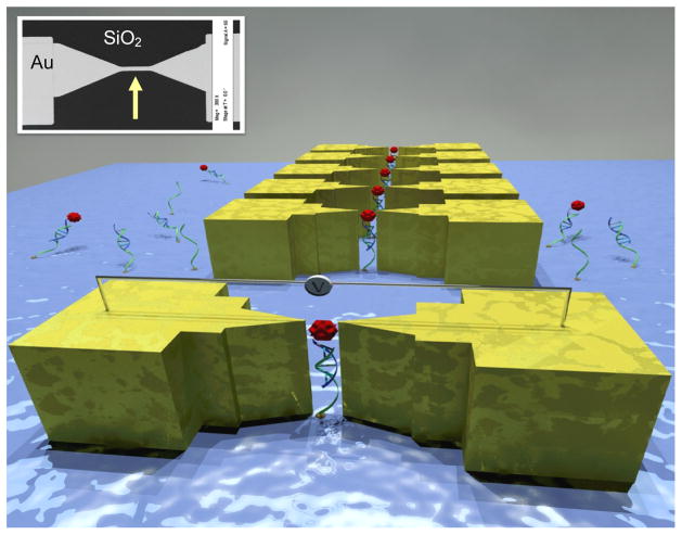
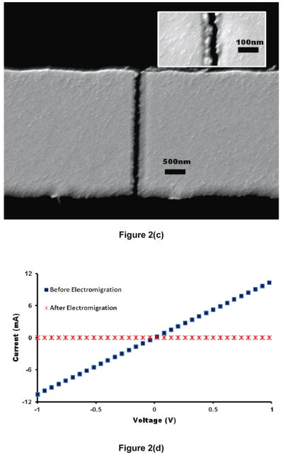
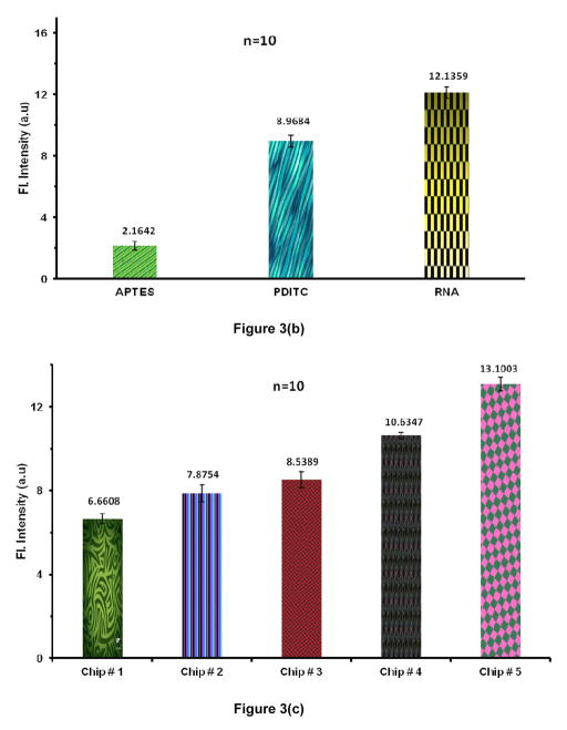
Epidermal growth factor receptor (EGFR) is a cell surface protein overexpressed in cancerous cells. It is known to be the most common oncogene. EGFR concentration also increases in the serum of cancer patients. The detection of small changes in the concentration of EGFR can be critical for early diagnosis, resulting in better treatment and improved survival rate of cancer patients. This article reports an RNA aptamer based approach to selectively capture EGFR protein and an electrical scheme for its detection. Pairs of gold electrodes with nanometer separation were made through confluence of focused ion beam scratching and electromigration. The aptamer was hybridized to a single stranded DNA molecule, which in turn was immobilized on the SiO(2) surface between the gold nanoelectrodes. The selectivity of the aptamer was demonstrated by using control chips with mutated non-selective aptamer and with no aptamer. Surface functionalization was characterized by optical detection and two orders of magnitude increase in direct current (DC) was measured when selective capture of EGFR occurred. This represents an electronic biosensor for the detection of proteins of interest for medical applications.


Figures

Figure 1
Figure 1
A 3D sketch of the device that demonstrates the mechanism of the electronic detection (not to scale). Inset shows an SEM micrograph of the thin Au line on SiO2 chip.
Figure 2
Figure 2
Characterization of the device (a) Representative I–V data after FIB scratching of metal line shows linear ohmic behavior. (b) A sudden drop in current confirms the complete breaking of metal line due to electromigration and results in nanogap break-junctions. (c) SEM micrograph showing a clean break between nanoelectrodes. Inset shows magnified view of a break-junction with a gap smaller than 30 nm. (d) Comparison of I–V data for a representative break-junction before and after the electromigration.
Figure 2
Figure 2
Characterization of the device (a) Representative I–V data after FIB scratching of metal line shows linear ohmic behavior. (b) A sudden drop in current confirms the complete breaking of metal line due to electromigration and results in nanogap break-junctions. (c) SEM micrograph showing a clean break between nanoelectrodes. Inset shows magnified view of a break-junction with a gap smaller than 30 nm. (d) Comparison of I–V data for a representative break-junction before and after the electromigration.
Figure 3
Figure 3
(a) Acridine Orange (AO) stain intensity measurements (background subtracted) depicts the presence of surface-bound DNA by comparing it with control chips. (n=10). (b) Fluorescence intensity measurements (background subtracted) using AO confirms the RNA hybridization to surface bound ssDNA when compared to control chips (n=10). (c) Sypro staining shows the selective capture of EGFR on functionalized SiO2. From L to R: Chip # 1: Control chip treated with blocking buffer (BB) but no aptamer; Chip # 2: Control chip with no BB treatment and no aptamer; Chip # 3: Control chip functionalized with mutant aptamer and treated with BB; Chip # 4: Functionalized chip with EGFR aptamer and captured protein after BB treatment; Chip # 5: Functionalized chip with EGFR aptamer and captured protein without using BB. (Error bars represent the standard deviation for n = 10)
Figure 3
Figure 3
(a) Acridine Orange (AO) stain intensity measurements (background subtracted) depicts the presence of surface-bound DNA by comparing it with control chips. (n=10). (b) Fluorescence intensity measurements (background subtracted) using AO confirms the RNA hybridization to surface bound ssDNA when compared to control chips (n=10). (c) Sypro staining shows the selective capture of EGFR on functionalized SiO2. From L to R: Chip # 1: Control chip treated with blocking buffer (BB) but no aptamer; Chip # 2: Control chip with no BB treatment and no aptamer; Chip # 3: Control chip functionalized with mutant aptamer and treated with BB; Chip # 4: Functionalized chip with EGFR aptamer and captured protein after BB treatment; Chip # 5: Functionalized chip with EGFR aptamer and captured protein without using BB. (Error bars represent the standard deviation for n = 10)
Figure 4
Figure 4
Comparison of I–V data for a representative break-junction before and after the surface modification and exposure to EGFR: (a) Anti-EGFR aptamer chip shows current increase due to the capture of EGFR bridging the nanogap; (b) Mutant aptamer functionalized control chip shows no change in conductivity, inset shows data for control chip with no aptamer and thus no change in conductivity. Both controls show no capture of EGFR.

References

    1. Klijn JGM, Berns P, Schmitz PIM, Foekens JA. The clinical significance of epidermal growth factor receptor (EGF-R) in human breast cancer: a review on 5232 patients. Endocrine reviews. 1992;13:3. - PubMed
    1. Lynch TJ, Bell DW, Sordella R, Gurubhagavatula S, Okimoto RA, Brannigan BW, Harris PL, Haserlat SM, Supko JG, Haluska FG. Activating mutations in the epidermal growth factor receptor underlying responsiveness of non-small-cell lung cancer to gefitinib. The New England journal of medicine. 2004;350:2129. - PubMed
    1. Kersemaekers AMF, Fleuren GJ, Kenter GG, Van den Broek L, Uljee SM, Hermans J, Van de Vijver MJ. Oncogene alterations in carcinomas of the uterine cervix: overexpression of the epidermal growth factor receptor is associated with poor prognosis. Clinical Cancer Research. 1999;5:577. - PubMed
    1. Mellon K, Wright C, Kelly P, Horne CH, Neal DE. Original Articles: Bladder Cancer: Long-Term Outcome Related to Epidermal Growth Factor Receptor Status in Bladder Cancer. The Journal of urology. 1995;153:919–25. - PubMed
    1. Inada S, Koto T, Futami K, Arima S, Iwashita A. Evaluation of malignancy and the prognosis of esophageal cancer based on an immunohistochemical study (p53, E-cadherin, epidermal growth factor receptor) Surgery today. 1999;29:493–503. - PubMed

Publication types

MeSH terms