. 2012 Jul 27;287(31):26342-51.
doi: 10.1074/jbc.M112.377168. Epub 2012 Jun 15.

A bipartite autoinhibitory region within the B-domain suppresses function in factor V

Affiliations

A bipartite autoinhibitory region within the B-domain suppresses function in factor V

Mettine H A Bos et al. J Biol Chem. .

Abstract

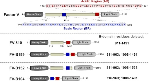
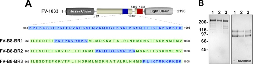
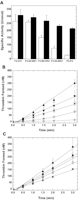
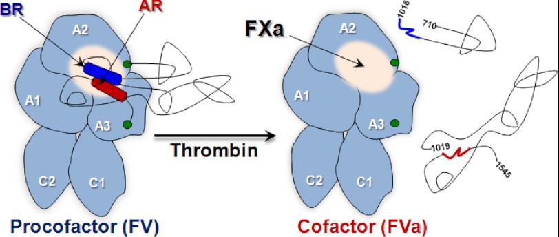
Activation of blood coagulation factor V (FV) is a key reaction of hemostasis. FV circulates in plasma as an inactive procofactor, and proteolytic removal of a large central B-domain converts it to an active cofactor (FVa) for factor Xa (FXa). Here we show that two short evolutionary conserved segments of the B-domain, together termed the procofactor regulatory region, serve an essential autoinhibitory function. This newly identified motif consists of a basic (963-1008) and an acidic (1493-1537) region and defines the minimal sequence requirements to maintain FV as a procofactor. Our data suggest that dismantling this autoinhibitory region via deletion or proteolysis is the driving force to unveil a high affinity binding site(s) for FXa. These findings document an unexpected sequence-specific role for the B-domain by negatively regulating FV function and preventing activity of the procofactor. These new mechanistic insights point to new ways in which the FV procofactor to cofactor transition could be modulated to alter hemostasis.


Figures

FIGURE 1.
FIGURE 1.
Recombinant FV derivatives. Top, the human FV B-domain (pale yellow) is defined by residues 710–1545, which are removed after thrombin-mediated proteolysis; cleavage sites are indicated below the B-domain. The blue box represents the BR (963–1008), and the red box represents the AR (1493–1537). Their sequences are provided. Recombinant FV derivative with variable B-domains used in the study are shown schematically (FV-810, FV-B199, FV-B152, and FV-B104). Because FV-B226, FV-B208, and FV-B199 are very similar, only FV-B199 is shown. B-domain residues deleted are provided on the right. The total lengths of the B-domains are as follows: FV-810, 155 residues; FV-B199, 199 residues; FV-B152, 152 residues; FV-B104, 104 residues. The full-length B-domain (710–1545) has 25 potential N-linked carbohydrate sites with a distribution as follows: 709–810, 6 sites; 811–962, 2 sites; 963–1008 (BR), 0 sites; 1009–1491, 16 sites; 1492–1545 (mostly AR), 1 site.
FIGURE 2.
FIGURE 2.
SDS-PAGE analysis. Purified proteins (5 μg/lane) before (left) and after treatment with thrombin (right) were subjected to SDS-PAGE under reducing conditions and visualized by staining with Coomassie Brilliant Blue R-250. Lane 1, FV-810; lane 2, FV-B226; lane 3, FV-B208; lane 4, FV-B199; lane 5, FV-B152; lane 6, FV-B104. The apparent molecular weights of the standards are indicated.
FIGURE 3.
FIGURE 3.
Characterization of rFV+BR derivatives. Panel A, specific clotting activities of recombinant FV/V(a) derivatives before (white bars) or after treatment with thrombin (black bars) were determined by a FV-specific PT-based clotting assay as described under “Experimental Procedures.” The data are the mean ± S.D. of at least three similar experiments. Panels B and C. Reaction mixtures containing 50 μm PCPS, 3.0 μm DAPA, 1.4 μm prothrombin, and 0.1 nm FV/V(a) derivatives before (panel B) or after treatment with thrombin (panel C) were incubated for 5 min at 25 °C. The reaction was initiated with 1.0 nm FXa, and thrombin generation was monitored as described under “Experimental Procedures.” The symbols represent the following: ▴, FV-810; ◊, FV-B226; ×, FV-B208; ○, FV-B199; ●, FV-B152; △, FV-B104; □, PD-FV; ■, rFVa. The lines represent a linear fit of the data, except for FV-B226, FV-B208, FV-B199, FV-B104, and PD-FV in panel B, which result from a polynomial fit. The data are the means ± S.D. of 2–4 similar experiments.
FIGURE 4.
FIGURE 4.
Direct binding measurements. Reaction mixtures containing 20 nm OG488-FXa and 50 μm PCPS were titrated with increasing concentrations of FV-810 (▴), FV-B226 (◊), FV-B208 (×), FV-B199 (○), PD-FV (□), and rFVa (▾; only shown in panel B) at 25 °C without prior treatment (panel A) or with proteins treated with thrombin before the experiment (panel B). The change in fluorescence anisotropy (Δr) was measured and analyzed as described in “Experimental Procedures.” The lines are drawn after analysis to independent, non-interacting sites. The data are representative of 2–4 similar experiments. The dissociation constants (Kd) for FV-810 in panel A was 1.1 ± 0.3 nm. We were not able to accurately determine Kd values for FV-B226, FV-B208, FV-B199, and PD-FV; they are estimated to be >50 nm. In panel B, the dissociation constants for the variants after thrombin treatment are as follows: FV-810, 1.3 ± 0.3 nm; FV-B226, 1.8 ± 0.5 nm; FV-B208, 2.1 ± 0.7 nm; FV-B199, 1.3 ± 0.3 nm; plasma-derived FVa, 1.5 ± 0.3 nm; rFVa, 1.8 ± 0.2 nm.
FIGURE 5.
FIGURE 5.
Schematic representation and SDS-PAGE analysis of FV-B8-BR variants. Panel A, FV-1033 is a procofactor-like FV derivative that harbors both a BR (blue box; sequence shown) and AR (red box) and has B-domain residues 1034–1491 deleted (26). With FV-1033 as a scaffold, portions of the BR were retained, and the remaining sequence was replaced with non-homologous segments of FVIII B-domain (green). The BR region retained for the different variants is as follows: 971–980 (FV-B8-BR1), 983–994 (FV-B8-BR2), and 997–1008 (FV-B8-BR3). Panel B, purified proteins (5 μg/lane) before (left) and after treatment with thrombin (right) were subjected to SDS-PAGE under reducing conditions and visualized by staining with Coomassie Brilliant Blue R-250. Lane 1, FV-B8-BR1; lane 2, FV-B8-BR2; lane 3, FV-B8-BR3. The apparent molecular weights of the standards are indicated.
FIGURE 6.
FIGURE 6.
Characterization of FV-B8-BR variants. Panel A, specific clotting activities of FV/FV(a) derivatives before (white bars) or after treatment with thrombin (black bars) were determined by a FV-specific PT-based clotting assay as described under “Experimental Procedures.” The data are the means ± S.D. of at least three experiments. FV-810 and PD-FV are for reference and are from Fig. 3A. Panels B and C, reaction mixtures containing 50 μm PCPS, 3.0 μm DAPA, 1.4 μm prothrombin, and 0.1 nm FV derivatives were incubated for 5 min at 25 °C. The reaction was initiated with 1.0 nm FXa, and thrombin generation was monitored as described under “Experimental procedures.” The symbols represent the following: FV-810 (▴), FV-B8-BR1 (▾), FV-B8-BR2 (*), FVB8-BR3 (⎔), PD-FV (□). The lines represent a linear fit of the data, except for FV-B8-BR3 and PD-FV in panel B, which results from a polynomial fit. The data are the means ± S.D. of 2–4 similar experiments.
FIGURE 7.
FIGURE 7.
Schematic of FV activation and functional landmarks. After thrombin processing to FVa, the B-domain (black lines) was released as two large fragments thereby exposing a FXa binding site. For FV, the BR (dark blue) and AR (dark red) are shown as cylinders; the putative FXa binding site is shown as a white sphere; green circles represent the end of the A2 and beginning of A3 domains.

References

    1. Khan A. R., James M. N. (1998) Molecular mechanisms for the conversion of zymogens to active proteolytic enzymes. Protein Sci. 7, 815–836 - PMC - PubMed
    1. Mann K. G., Kalafatis M. (2003) Factor V. A combination of Dr. Jekyll and Mr. Hyde. Blood 101, 20–30 - PubMed
    1. Camire R. M., Bos M. H. (2009) The molecular basis of factor V and VIII procofactor activation. J. Thromb. Haemost. 7, 1951–1961 - PMC - PubMed
    1. Mann K. G., Nesheim M. E., Church W. R., Haley P., Krishnaswamy S. (1990) Surface-dependent reactions of the vitamin K-dependent enzyme complexes. Blood 76, 1–16 - PubMed
    1. Nesheim M. E., Mann K. G. (1979) Thrombin-catalyzed activation of single chain bovine factor V. J. Biol. Chem. 254, 1326–1334 - PubMed

Publication types

LinkOut - more resources