Expression of apoptosis regulatory markers in the skin of advanced hepatitis-C virus liver patients
- PMID: 22707768
- PMCID: PMC3371520
- DOI: 10.4103/0019-5154.96189
Expression of apoptosis regulatory markers in the skin of advanced hepatitis-C virus liver patients
Abstract
Background: Hepatitis-C virus (HCV) infection is considered a major worldwide public health problem with a global prevalence. Maintenance of skin homeostasis requires a delicate balance between proliferation, differentiation, and apoptosis. Meanwhile, it is unclear if there is an altered keratinocyte proliferation/apoptosis balance in advanced liver disease with HCV infection.
Aim: This work aimed to evaluate the epidermal thickness and changes in the expression of apoptosis regulatory markers as well as apoptotic index in skin samples of advanced HCV liver patients compared to normal controls.
Materials and methods: Twenty biopsies were taken from apparently normal skin of advanced HCV liver disease patients, as well as five healthy control subjects. These specimens were used for histometric epidermal measurement, immunohistochemical staining of apoptosis regulatory proteins (Bax, Fas, p53, Caspase-3, Bcl-2, Bcl-xL) as well as the TUNEL technique for detection of apoptotic cells.
Results: The mean epidermal thickness was significantly lower than the control group (P=0.000). There were significant overexpression of pro-apoptotic markers (Bax, Fas, P53, and Caspase-3) in patients (P=0.03, 0.03, 0.003, 0.003 respectively), with increased apoptotic index in HCV liver patients (P=0.002) when compared to normal controls. On the other hand, no statistically significant difference were encountered in the expression of antiapoptotic markers (Bcl-2, Bcl-xL) in HCV patients when compared to normal controls (P=0.5, 0.9, respectively).
Conclusion: These findings suggest that an alteration in the proliferation/apoptosis balance is present in the skin of HCV liver patients.
Keywords: Apoptosis; Bcl-2; epidermal thickness; hepatitis-C virus; liver disease.
Conflict of interest statement
Figures


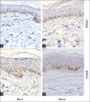
Significant increase of cytoplasmic expression of Bax staining in basal and squamous cell layers in patients (a) when compared to that confined to the basal cell layer of controls (b) (Immunohistochemical, ×400).
Significant increase of membranous expression of Fas staining in basal and lower 2/3 of squamous cell layers in patients (c) when compared to that confined to the basal cell layer of controls (d) (Immunohistochemical, ×400).
Significant increase of nuclear expression of P53 staining in basal and lower 2/3 of squamous cell layers in patients (e) when compared to that confined to the basal cell layer of controls (f) (Immunohistochemical, ×200).
Significant increase of cytoplasmic expression of Caspase 3 staining in basal and squamous cell layers in patients (g) when compared to minimal staining confined to the basal cell layer of controls (h) (Immunohistochemical, ×400)

Insiginificant lower cytoplasmic expression of Bcl-2 staining, mainly confined to the basal cell layer, in patients (a) when compared to controls (b).
Insiginificant lower perinuclear cytoplasmic expression of Bcl-xL staining, mainly confined to the basal cell layer, in patients (c) when compared to that confined to the basal and supra basal cell layer of controls (d) (Immunohistochemical, ×400).

Similar articles
-
Expression of apoptosis regulatory proteins p53 and Bcl-2 in skin of patients with chronic renal failure on maintenance haemodialysis.J Eur Acad Dermatol Venereol. 2007 Jul;21(6):795-801. doi: 10.1111/j.1468-3083.2006.02090.x. J Eur Acad Dermatol Venereol. 2007. PMID: 17567310
-
Potential role of bcl-2 and bax mRNA and protein expression in chronic hepatitis type B and C: a clinicopathologic study.Mod Pathol. 2003 Dec;16(12):1273-88. doi: 10.1097/01.MP.0000097367.56816.5E. Mod Pathol. 2003. PMID: 14681329
-
Evaluation of apoptosis regulatory proteins in response to PUVA therapy for psoriasis.Photodermatol Photoimmunol Photomed. 2013 Feb;29(1):18-26. doi: 10.1111/phpp.12012. Photodermatol Photoimmunol Photomed. 2013. PMID: 23281693 Clinical Trial.
-
Fas-mediated hepatocyte apoptosis is increased by hepatitis C virus infection and alcohol consumption, and may be associated with hepatic fibrosis: mechanisms of liver cell injury in chronic hepatitis C virus infection.J Viral Hepat. 2001 Nov;8(6):406-13. doi: 10.1046/j.1365-2893.2001.00316.x. J Viral Hepat. 2001. PMID: 11703571
-
Characterization of chronic HCV infection-induced apoptosis.Comp Hepatol. 2011 Jul 23;10(1):4. doi: 10.1186/1476-5926-10-4. Comp Hepatol. 2011. PMID: 21781333 Free PMC article.
Cited by
-
Increased Spontaneous Programmed Cell Death Is Associated with Impaired Cytokine Secretion in Peripheral Blood Mononuclear Cells from Hepatitis C Virus-Positive Patients.Viral Immunol. 2017 May;30(4):283-287. doi: 10.1089/vim.2016.0166. Epub 2017 Mar 17. Viral Immunol. 2017. PMID: 28304236 Free PMC article.
-
Effect of the new synthetic vitamin E derivative ETS-GS on radiation enterocolitis symptoms in a rat model.Oncol Lett. 2013 Nov;6(5):1229-1233. doi: 10.3892/ol.2013.1581. Epub 2013 Sep 12. Oncol Lett. 2013. PMID: 24179500 Free PMC article.
-
Altered of apoptotic markers of both extrinsic and intrinsic pathways induced by hepatitis C virus infection in peripheral blood mononuclear cells.Virol J. 2012 Dec 20;9:314. doi: 10.1186/1743-422X-9-314. Virol J. 2012. PMID: 23256595 Free PMC article.
-
Apoptosis and clinical severity in patients with psoriasis and HCV infection.Indian J Dermatol. 2014 May;59(3):230-6. doi: 10.4103/0019-5154.131377. Indian J Dermatol. 2014. PMID: 24891650 Free PMC article.
References
-
- Chapman RW, Collier JD, Hayes PC. Liver and biliary tract disease. In: Nicholas A, Nicki R, Brian R, editors. Davidsons's Principles and Practice of Medicine. 20th ed. USA: Elsevier Science; 2006. pp. 935–98.
-
- Azfar NA, Zaman T, Rashid T, Jahangir M. Cutaneous manifestations in patients of hepatitis C. J Pak Med Assoc Dermatol. 2008;18:138–43.
-
- Hepatitis C. Geneva: WHO; C2004, 2008. [accessed on 2008 Nov 15]. World Health Organisation (WHO) Available from: http://www.who.int/mediacentre/factssheets/fs164/en/
-
- Mohamed MK. Epidemiology of HCV in Egypt. Afro-Arab Liver Jr. 2004;3:41–52.
-
- Nafeh MA, Medhat A, Shehata M, Mikhail NN, Swifee Y, Abdel-Hamid M, et al. Hepatitis C in a community in upper Egypt: Cross-sectional survey. Am J Trop Med Hyg. 2004;63:236–41. - PubMed
LinkOut - more resources
Full Text Sources
Research Materials
Miscellaneous
