

Angiosarcomas of the bilateral breast and heart: which one is the primary site?
- PMID: 22707897
- PMCID: PMC3372809
- DOI: 10.3904/kjim.2012.27.2.224
Angiosarcomas of the bilateral breast and heart: which one is the primary site?
Abstract
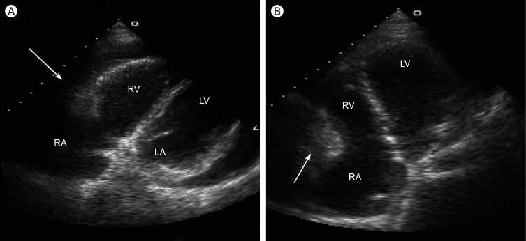
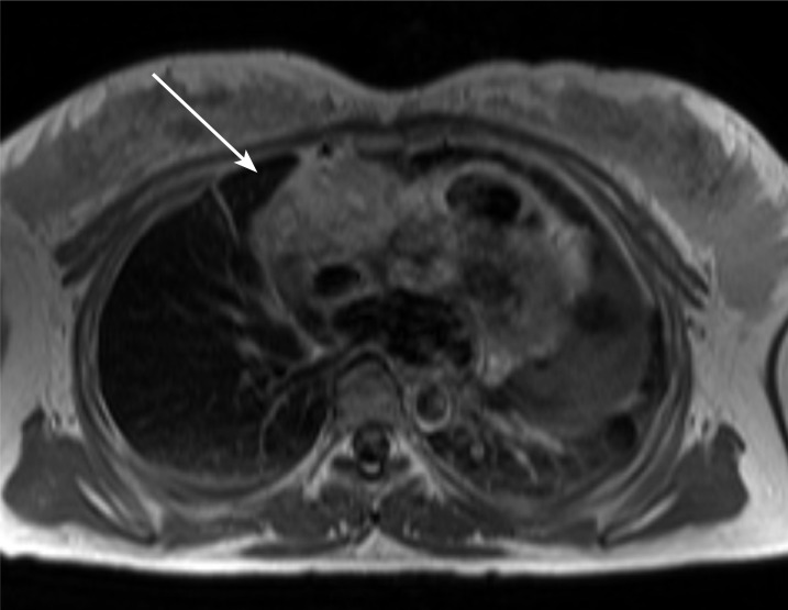
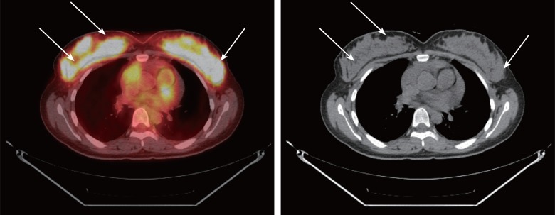
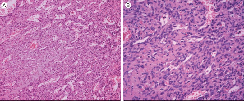
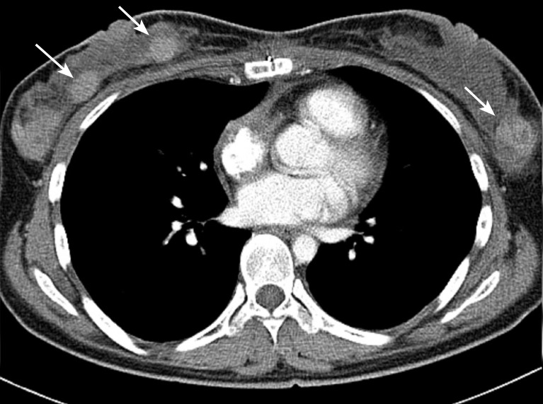
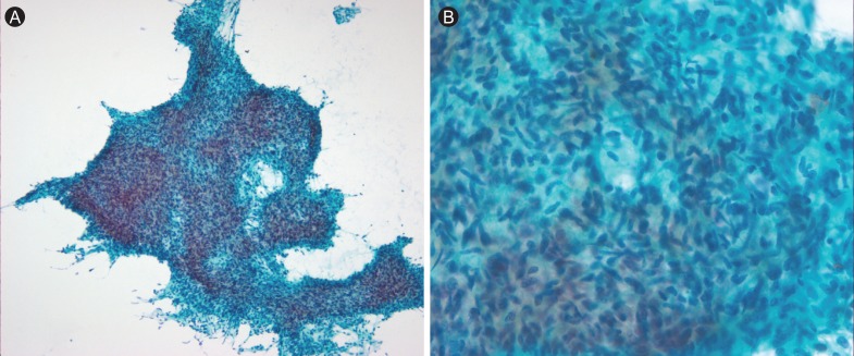
A 29-year-old pregnant woman with recurrent pericardial effusion and a cardiac tumor, diagnosed as an angiosarcoma, was treated with surgical resection of the tumor followed by radiotherapy. Immediately after completion of radiotherapy, she developed bilateral breast masses, which were also confirmed as angiosarcomas. We thought this might be the first case of bilateral angiosarcoma of the breast metastasizing to heart mimicking a primary cardiac angiosarcoma, although we could not conclude with certainty that angiosarcoma of the heart was not the primary site.
Keywords: Breast; Heart; Hemangiosarcoma.
Conflict of interest statement
No potential conflict of interest relevant to this article was reported.
Figures
Similar articles
-
Angiosarcoma presenting as syncope.Asian Cardiovasc Thorac Ann. 2008 Apr;16(2):154-6. doi: 10.1177/021849230801600216. Asian Cardiovasc Thorac Ann. 2008. PMID: 18381877
-
Hemorrhagic tamponade due to cardiac angiosarcoma.South Med J. 2010 Oct;103(10):1055-7. doi: 10.1097/SMJ.0b013e3181ecfb64. South Med J. 2010. PMID: 20802380
-
Angiosarcoma in a pregnant woman presenting with pericardial tamponade--a case report and review of the literature.Angiology. 2006 Mar-Apr;57(2):251-7. doi: 10.1177/000331970605700219. Angiology. 2006. PMID: 16518537
-
Primary angiosarcoma of the pericardium: case report and review of the literature.Kardiol Pol. 2010 Jul;68(7):802-5. Kardiol Pol. 2010. PMID: 20648441 Review.
-
[Primary angiosarcoma of the heart. A report of a new case and a review of the literature].Rev Clin Esp. 1992 Apr;190(6):302-4. Rev Clin Esp. 1992. PMID: 1598428 Review. Spanish.
Cited by
-
Bilateral breast masses with a rare etiology.Case Rep Oncol Med. 2013;2013:412368. doi: 10.1155/2013/412368. Epub 2013 Aug 28. Case Rep Oncol Med. 2013. PMID: 24066248 Free PMC article.
-
Primary Angiosarcoma of the Breast: A Case Report and Review of Literature.World J Oncol. 2014 Jun;5(3):144-148. doi: 10.14740/wjon809w. Epub 2014 Jun 25. World J Oncol. 2014. PMID: 29147394 Free PMC article.
-
Primary and secondary angiosarcoma of the breast.Gland Surg. 2014 Feb;3(1):28-34. doi: 10.3978/j.issn.2227-684X.2013.12.03. Gland Surg. 2014. PMID: 25083491 Free PMC article. Review.
References
-
- Chen KT, Kirkegaard DD, Bocian JJ. Angiosarcoma of the breast. Cancer. 1980;46:368–371. - PubMed
-
- Talbot SM, Taub RN, Keohan ML, Edwards N, Galantowicz ME, Schulman LL. Combined heart and lung transplantation for unresectable primary cardiac sarcoma. J Thorac Cardiovasc Surg. 2002;124:1145–1148. - PubMed
-
- Agarwal PK, Mehrotra R. Haemangiosarcoma of the breast. Indian J Cancer. 1977;14:182–185. - PubMed
-
- West JG, Qureshi A, West JE, et al. Risk of angiosarcoma following breast conservation: a clinical alert. Breast J. 2005;11:115–123. - PubMed
-
- Kiluk JV, Yeh KA. Primary angiosarcoma of the breast. Breast J. 2005;11:517–518. - PubMed
Publication types
MeSH terms
Supplementary concepts
LinkOut - more resources
Full Text Sources
Medical