. 2012 Jul;3(Suppl 1):155-65.
doi: 10.1007/s12975-012-0166-9. Epub 2012 Apr 14.

Heparin reduces neuroinflammation and transsynaptic neuronal apoptosis in a model of subarachnoid hemorrhage

Heparin reduces neuroinflammation and transsynaptic neuronal apoptosis in a model of subarachnoid hemorrhage

J Marc Simard et al. Transl Stroke Res. 2012 Jul.

Abstract

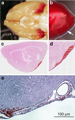
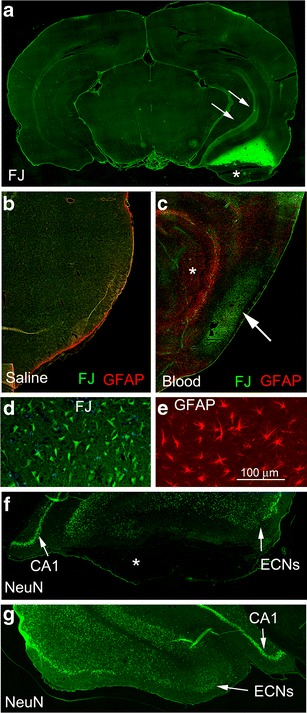
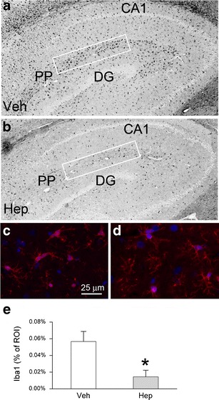
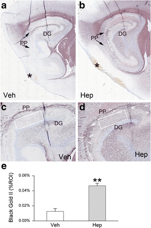
Subarachnoid hemorrhage (SAH) can lead to disabling motor, cognitive, and neuropsychological abnormalities. Part of the secondary injury to cerebral tissues associated with SAH is attributable to the neuroinflammatory response induced by blood. Heparin is a pleiotropic compound that reduces inflammatory responses in conditions outside the central nervous system. Using a model of SAH devoid of global insult, we evaluated the effect of delayed intravenous (IV) infusion of heparin, at a dose that does not produce therapeutic anticoagulation, on neuroinflammation, myelin preservation, and apoptosis. Adult male rats underwent bilateral stereotactic injections of autologous blood (50 μL) into the subarachnoid space of the entorhinal cortex. The rats were implanted with mini-osmotic pumps that delivered either vehicle or unfractionated heparin (10 U/kg/h IV) beginning 12 h after SAH. No mechanical or hemorrhagic injury was observed in the hippocampus. In vehicle controls assessed at 48 h, SAH was associated with robust neuroinflammation in the adjacent cortex [neutrophils, activated phagocytic microglia, nuclear factor-kappa B, tumor necrosis factor-alpha, and interleukin-1beta] and neurodegeneration (Fluoro-Jade C staining and loss of NeuN). In the hippocampus, a muted neuroinflammatory response was indicated by Iba1-positive, ED1-negative microglia exhibiting an activated morphology. The perforant pathway showed Fluoro-Jade C staining and demyelination, and granule cells of the dentate gyrus had pyknotic nuclei, labeled with Fluoro-Jade C and showed upregulation of cleaved caspase-3, consistent with transsynaptic apoptosis. Administration of heparin significantly reduced neuroinflammation, demyelination, and transsynaptic apoptosis. We conclude that delayed IV infusion of low-dose unfractionated heparin may attenuate adverse neuroinflammatory effects of SAH.


Figures

Fig. 1
Fig. 1
SAH involving the entorhinal cortex. a Image of the base of the brain showing SAH involving the entorhinal cortex bilaterally; dotted line location of parasagittal sections. b Parasagittal section of the brain stained with TTC showing the absence of infarction in the region of SAH (arrow). c–e Parasagittal sections of the brain in the region of SAH stained with H&E showing that the hemorrhage is localized largely in the subarachnoid space and that subarachnoid vessels show no evidence of vasoconstriction or vasospasm. All tissues were harvested 48 h after injury; the images shown are representative of findings in 28 rats (a), 3 rats (b), or 5 rats (ce)
Fig. 2
Fig. 2
Cortical neurodegeneration following SAH. a Coronal section from a rat with unilateral injection of 50 μL of autologous blood into the subarachnoid space of the entorhinal cortex, stained with Fluoro-Jade C (FJ); note the bright staining above the background in the cortex and underlying white matter near the hemorrhage, along with the bright staining of the perforant pathway ipsilateral (arrows) but not contralateral to the hemorrhage; asterisk denotes hemorrhage. b–e Parasagittal sections of rat brains in the region of injection of saline (b) or blood (ce), stained with Fluoro-Jade C (bd) and immunolabeled for GFAP (b, c, e) showing degeneration involving neuron-like cells in the cortex beneath the SAH (c, d), but not with saline (b) and reactive astrocytes surrounding the region that stains for Fluoro-Jade C (c, e). f, g Coronal sections from a rat with unilateral injection of 50 μL of autologous blood into the subarachnoid space of the entorhinal cortex, immunolabeled for NeuN, showing the entorhinal cortex ipsilateral (f) and contralateral (g) to the hemorrhage; note the loss of ipsilateral entorhinal cortex neurons (ECNs) throughout the region, except laterally, and the preservation of neurons in the ipsilateral CA1 region and in all regions contralaterally (g); asterisk denotes hemorrhage; same rat as in a. All tissues were harvested 48 h after injury; the images shown are representative of findings in five rats (be) or three rats (a, f, g)
Fig. 3
Fig. 3
Cortical neuroinflammation following SAH. a, b Parasagittal section immunolabeled for NF-κB (p65), shown at low (a) and high (b) magnification, demonstrating (1) prominent expression of p65 subpially near the hemorrhage (asterisk) that tapers off with distance (a); (2) involvement of individual cells as well as elongated structures consistent with microvessels (arrows) (b); the image shown in a is an “inverse” epifluorescence image. c Parasagittal section immunolabeled for myeloperoxidase (MPO) to identify neutrophils (note also the polymorphic nuclei) and ED1 to identify macrophages and phagocytic, activated microglia, showing tissue infiltration beneath the pia (dotted line). d Parasagittal section immunolabeled for Iba1, to identify microglia, showing (1) large rounded cells (a and inset a) beneath the pia (dotted line), consistent with the phagocytic, activated microglia labeled with ED1 in c; (2) small cells with complex processes further away from the hemorrhage (b and inset b). All tissues were harvested 48 h after injury; the images shown are representative of findings in five rats
Fig. 4
Fig. 4
Heparin reduces cortical neuroinflammation (microglial activation) following SAH. a–d Parasagittal sections immunolabeled for myeloperoxidase (MPO) to identify neutrophils (a, b) or ED1 to identify macrophages and phagocytic, activated microglia (c, d) from rats treated with vehicle (a, c) or heparin (b, d); the advancing front of neutrophils, depicted by arrows (a, b), was similar without and with heparin; the advancing front of ED1-positive macrophages and microglia, depicted by arrows (c, d), was reduced by heparin; asterisk denotes the location of the SAH; the images shown are “inverse” epifluorescence images. e Bar graphs showing counts of MPO-positive cells, ED1-positive cells, and upregulation of p65 in rats treated with vehicle (Veh) or heparin (Hep); *P < 0.05; **P < 0.01; five rats per group
Fig. 5
Fig. 5
Heparin reduces cortical neuroinflammation (TNFα and IL-1β) following SAH. a–d Parasagittal sections immunolabeled for TNFα (a, b) or IL-1β (c, d) from rats treated with vehicle (a, c) or heparin (b, d); the advancing front of TNFα, depicted by arrows (a, b), was reduced by heparin; asterisk denotes the location of the SAH; the images shown in a and b are “inverse” epifluorescence images. e Bar graphs showing the percentage of the ROI immunolabeled for TNFα or IL-1β, as well as the number of cells immunolabeled for IL-1β, as indicated, in rats treated with vehicle (Veh) or heparin (Hep); *P < 0.05; **P < 0.01; five rats per group
Fig. 6
Fig. 6
Heparin reduces hippocampal neuroinflammation following SAH. a–d Parasagittal sections immunolabeled for Iba1, shown at low (a, b) and high (c, d) magnification, from rats treated with vehicle (a, c) or heparin (b, d); note the “plump” morphology of microglia in the controls (c), which is absent with heparin (d); CA1 cornu ammonis region 1, PP perforant pathway, DG dentate gurus; the ROI analyzed are depicted by rectangles; the images shown in a and b are “inverse” epifluorescence images. e Bar graph quantifying Iba1 in the ROI in rats treated with vehicle (Veh) or heparin (Hep); *P < 0.05; five rats per group
Fig. 7
Fig. 7
Heparin reduces demyelination of the perforant pathway following SAH. a–d Parasagittal sections stained with Black Gold II, shown at low (a, b) and high (c, d) magnification, from rats treated with vehicle (a, c) or heparin (b, d); PP perforant pathway, DG dentate gyrus; the ROI analyzed is depicted by the rectangle. e Bar graph quantifying Black Gold II-stained myelin in the ROI in rats treated with vehicle (Veh) or heparin (Hep); **P < 0.01; five rats per group
Fig. 8
Fig. 8
Heparin reduces apoptosis of granule cells of the dentate gyrus following SAH. a–d Parasagittal sections of the dentate gyrus immunolabeled for cleaved caspase-3 (a, b) or stained with Fluoro-Jade C (c, d) from rats treated with vehicle (a, c) or heparin (b, d). e Granule cells of the dentate gyrus from a vehicle-treated rat, double stained with DAPI and with Fluoro-Jade C (FJ), as indicated, showing pyknotic nuclei of apoptotic cells (arrows); superimposed images are also shown. f Bar graph quantifying Fluoro-Jade C-stained granule cells in the dentate gyrus of naïve (uninjured) rats (CTR), rats with bilateral SAH treated with vehicle (Veh) or heparin (Hep); **P < 0.01; three naïve rats and eight rats per group with SAH

References

    1. Amiconi G, Zolla L, Vecchini P, et al. The effect of macromolecular polyanions on the functional properties of human hemoglobin. Eur J Biochem. 1977;76:339–343. doi: 10.1111/j.1432-1033.1977.tb11601.x. - DOI - PubMed
    1. Ayer R, Jadhav V, Sugawara T, et al. The neuroprotective effects of cyclooxygenase-2 inhibition in a mouse model of aneurysmal subarachnoid hemorrhage. Acta Neurochir Suppl. 2011;111:145–149. doi: 10.1007/978-3-7091-0693-8_24. - DOI - PubMed
    1. Bauer J, Sminia T, Wouterlood FG, et al. Phagocytic activity of macrophages and microglial cells during the course of acute and chronic relapsing experimental autoimmune encephalomyelitis. J Neurosci Res. 1994;38:365–375. doi: 10.1002/jnr.490380402. - DOI - PubMed
    1. Berry CN, Girard D, Lochot S, et al. Antithrombotic actions of argatroban in rat models of venous, ‘mixed’ and arterial thrombosis, and its effects on the tail transection bleeding time. Br J Pharmacol. 1994;113:1209–1214. - PMC - PubMed
    1. Bhardwaj A. Molecular targets for ameliorating early brain injury post subarachnoid hemorrhage: a new focus. Crit Care Med. 2010;38:727–728. doi: 10.1097/CCM.0b013e3181c0de31. - DOI - PubMed