. 2011 Jun 1;1(3):209-222.
doi: 10.1007/s13346-011-0022-6.

Development and Characterization of a Vaginal Film Containing Dapivirine, a Non- nucleoside Reverse Transcriptase Inhibitor (NNRTI), for prevention of HIV-1 sexual transmission

Affiliations

Development and Characterization of a Vaginal Film Containing Dapivirine, a Non- nucleoside Reverse Transcriptase Inhibitor (NNRTI), for prevention of HIV-1 sexual transmission

Ayman Akil et al. Drug Deliv Transl Res. .

Abstract

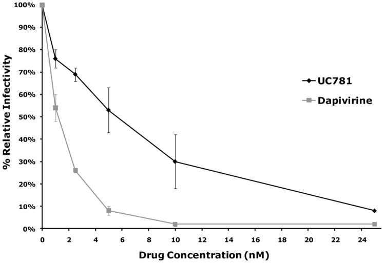
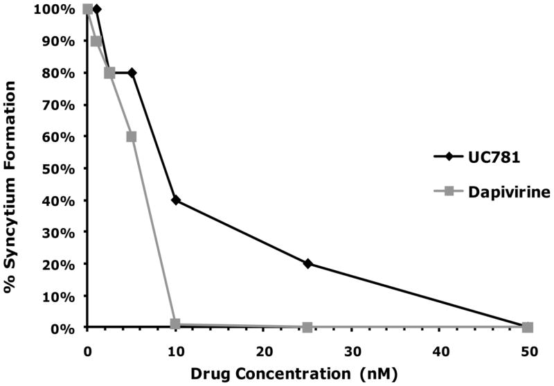
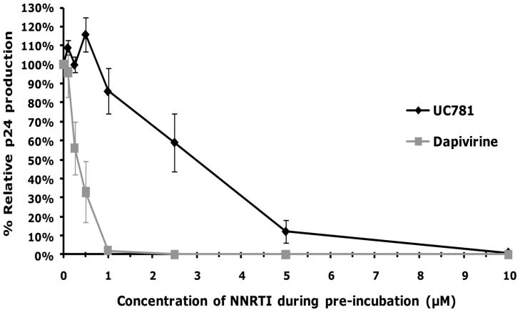
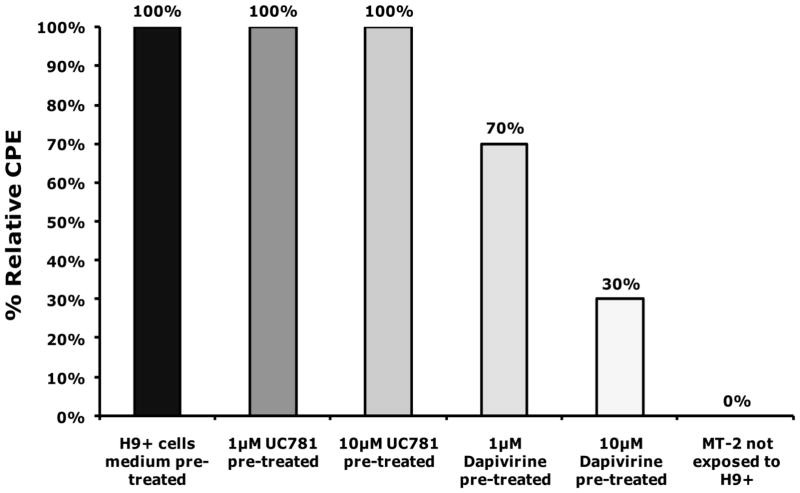
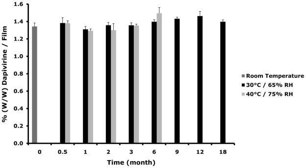
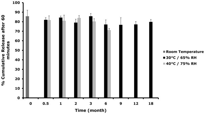
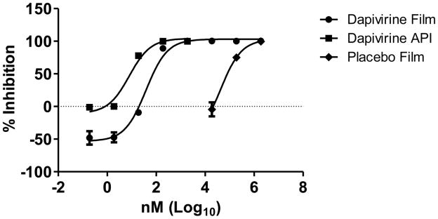
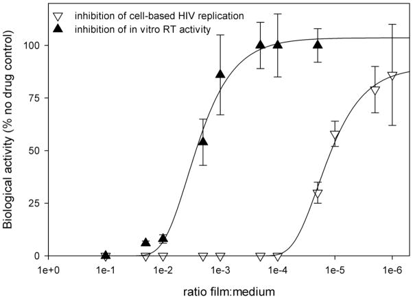
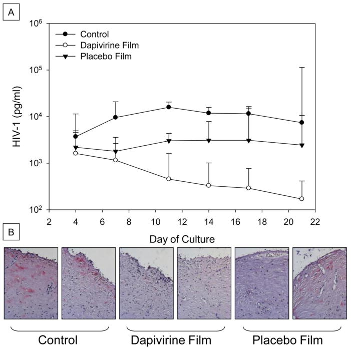
Dapivirine, a non-nucleoside reverse transcriptase inhibitor, is a potent and promising anti-HIV molecule. It is currently being investigated for use as a vaginal microbicide in two dosage forms, a semi-solid gel and a silicone elastomer ring. Quick-dissolving films are promising and attractive dosage forms that may provide an alternative platform for the vaginal delivery of microbicide drug candidates. Vaginal films may provide advantages such as discreet use, no product leakage during use, lack of requirement for an applicator for insertion, rapid drug release and minimal packaging and reduced wastage. Within this study the in vitro bioactivity of dapivirine as compared to the NNRTI UC781 was further established and a quick dissolve film was developed for vaginal application of dapivirine for prevention of HIV infection. The developed film was characterized with respect to its physical and chemical attributes including water content, mechanical strength, drug release profile, permeability, compatibility with lactobacilli and bioactivity. The anti-HIV activity of the formulated dapivirine film was confirmed in in vitro and ex vivo models. Importantly the physical and chemical properties of the film as well as its bioactivity were maintained for a period of 18 months. In conclusion, a vaginal film containing dapivirine was developed and characterized. The film was shown to prevent HIV-1 infection in vitro and ex vivo and have acceptable characteristics which make this film a promising candidate for testing as vaginal microbicide.


Figures

Figure 1
Figure 1
(A) P4R5 cells were exposed to varying concentrations of dapivirine or UC781, and then immediately inoculated with HIV-1 in the continued presence of the drug. The extent of infection was assessed after 48h using a fluorescence-based β-galactosidase detection assay using 4-methyllumbelliferyl-β-D-galactopyranoside. Dapivirine showed greater potency than UC781 in single HIV-1 replication cycle assays using the P4R5 indicator cell line. Dapivirine EC50 is 1 nM and UC781 EC50 is 5 nM. (B) P4R5 cells were exposed to varying concentrations of dapivirine or UC781 for 18h. The medium was removed and the cells were extensively washed and inoculated with HIV-1 virus in the absence of exogenous drug. The extent of infection was assessed after 48h. Both dapivirine and UC781 were able to establish a protective barrier to infection in P4R5 cells following drug pretreatment and subsequent exposure to HIV-1 in the absence of exogenous drug. Dapivirine showed greater potency than UC781 in this context.
Figure 1
Figure 1
(A) P4R5 cells were exposed to varying concentrations of dapivirine or UC781, and then immediately inoculated with HIV-1 in the continued presence of the drug. The extent of infection was assessed after 48h using a fluorescence-based β-galactosidase detection assay using 4-methyllumbelliferyl-β-D-galactopyranoside. Dapivirine showed greater potency than UC781 in single HIV-1 replication cycle assays using the P4R5 indicator cell line. Dapivirine EC50 is 1 nM and UC781 EC50 is 5 nM. (B) P4R5 cells were exposed to varying concentrations of dapivirine or UC781 for 18h. The medium was removed and the cells were extensively washed and inoculated with HIV-1 virus in the absence of exogenous drug. The extent of infection was assessed after 48h. Both dapivirine and UC781 were able to establish a protective barrier to infection in P4R5 cells following drug pretreatment and subsequent exposure to HIV-1 in the absence of exogenous drug. Dapivirine showed greater potency than UC781 in this context.
Figure 2
Figure 2
(A) MT-2 cells were exposed to varying concentrations of dapivirine or UC781, and then immediately inoculated with HIV-1 (25 ng HIV-1 p24/well) in the continued presence of the drug. The extent of infection was evaluated four days after virus addition by microscopic observation of HIV-1 induced syncytium formation. Dapivirine showed greater direct antiviral potency than UC781. Dapivirine EC50 is 5 nM and UC781 EC50 is 10 nM. (B) Uninfected MT-2 cells were incubated with various concentrations of dapivirine or UC781 for 18 hours. Exogenous drug was removed by extensive washing of the cells with culture medium. The washed cells were infected with equivalent infectious doses of HIV-1 (25 ng HIV-1 p24/well). The extent of viral infection was determined four days later by microscopic observation of HIV-1 induced syncytium formation and validated by measuring the level of HIV-1 p24 antigen in the cell-free culture supernatants. Both dapivirine and UC781 were able to establish a protective barrier to infection in MT-2 cells following drug pretreatment and subsequent exposure to HIV-1 in the absence of exogenous drug. Dapivirine again showed greater potency than UC781.
Figure 2
Figure 2
(A) MT-2 cells were exposed to varying concentrations of dapivirine or UC781, and then immediately inoculated with HIV-1 (25 ng HIV-1 p24/well) in the continued presence of the drug. The extent of infection was evaluated four days after virus addition by microscopic observation of HIV-1 induced syncytium formation. Dapivirine showed greater direct antiviral potency than UC781. Dapivirine EC50 is 5 nM and UC781 EC50 is 10 nM. (B) Uninfected MT-2 cells were incubated with various concentrations of dapivirine or UC781 for 18 hours. Exogenous drug was removed by extensive washing of the cells with culture medium. The washed cells were infected with equivalent infectious doses of HIV-1 (25 ng HIV-1 p24/well). The extent of viral infection was determined four days later by microscopic observation of HIV-1 induced syncytium formation and validated by measuring the level of HIV-1 p24 antigen in the cell-free culture supernatants. Both dapivirine and UC781 were able to establish a protective barrier to infection in MT-2 cells following drug pretreatment and subsequent exposure to HIV-1 in the absence of exogenous drug. Dapivirine again showed greater potency than UC781.
Figure 3
Figure 3
H9+ cells were incubated for 18 hrs in the absence or presence of 1 μM or 10 μM of dapivirine or UC781. The cells were washed extensively with culture medium to remove exogenous drug and then were co-cultured with uninfected MT-2 cells. The extent of viral infection of the MT-2 cells was determined after three days of co-culture by visual comparison of CPE relative to cells that were not treated with either compound. Dapivirine pre-treatment of MT-2/H9+ cell co-cultures resulted in 70% reduction in CPE levels compared to control MT-2/H9+ co-cultures. Dapivirine can inhibit HIV-1 cell to cell transmission.
Figure 4
Figure 4
Dapivirine was mixed with the film excipient mixture (composed of propylene glycol PVA, HPMC, and PEG8000) and stored at 30°C/65% relative humidity (RH), 40°C/75% RH or 50°C. Dapivirine drug content was determined using a UPLC method. No loss of dapivirine was observed after 14 days storage in the three storage conditions.
Figure 5
Figure 5
Dapivirine film (1.25 mg/film) dissolution was tested in standard class IV USP method. The medium used was a 1% Cremophor aqueous solution. Dapivirine was rapidly released from the film. 50% of dapivirine was released from the film in less than 10 minutes. Data presented as mean ± SD (n = 3).
Figure 6
Figure 6
Dapivirine film (1.25 mg/film) was stored for 18 month in 30°C/65% RH and for 6 in 40°C/75% RH. Several physical and chemical characteristics were monitored over the time course of the stability study including weight, drug and dissolution. (A) No change was observed in dapivirine film weight over the time course of stability testing. Data presented as mean ± SD. (B) Dapivirine film amount remained stable in the film at the different conditions tested. Data presented as mean ± SD. (C) Four films were tested per each time point. Dissolution testing over the study period showed no change in release values (cumulative release of dapivirine after 60 minutes). Data presented as mean ± SD. Three films were tested per each time point.
Figure 6
Figure 6
Dapivirine film (1.25 mg/film) was stored for 18 month in 30°C/65% RH and for 6 in 40°C/75% RH. Several physical and chemical characteristics were monitored over the time course of the stability study including weight, drug and dissolution. (A) No change was observed in dapivirine film weight over the time course of stability testing. Data presented as mean ± SD. (B) Dapivirine film amount remained stable in the film at the different conditions tested. Data presented as mean ± SD. (C) Four films were tested per each time point. Dissolution testing over the study period showed no change in release values (cumulative release of dapivirine after 60 minutes). Data presented as mean ± SD. Three films were tested per each time point.
Figure 7
Figure 7
Dapivirine and placebo films were dissolved in 2 ml saline and serial dilutions made. The active pharmaceutical ingredient (API) was dissolved in DMSO and serial dilutions made. The dilutions were applied to the TZM-bl cells in triplicate with HIV-1BaL. After 48 hours, HIV-1 infection was measured by adding BrightGlo (Promega) to the plates and reading for luminescent activity. The data show the API with an IC50 of 7.9 nM and the Dapivirine film with an IC50 of 38.3 nM. Minimal anti-HIV-1 activity was associated with the film.
Figure 8
Figure 8
Weighed pieces of dapivirine films were dissolved in either DMEM (for cell-based assays) or 1X RT-buffer (for in vitro RT assays) at 1:10 w/v. Multiple dilutions of the starting solutions were then prepared and used. For the RT assay, inhibition of the RNA-dependent DNA polymerase activity of HIV-1 RT was determined using poly(rA)-oligo(dT) and [3H-TTP]. Cell-based single HIV-1 replication cycle assays for direct antiviral activity of the film formulations used P4R5 indicators cells as described above. Inhibition of cell-based HIV-1 replication by dapivirine film (0.5 mg/film) is over 200-fold more potent than inhibition of RT activity.
Figure 9
Figure 9
Dapivirine and placebo films were dissolved in 2 ml of medium and 100 μl was applied to the apical surface of the designated explants while HIV-1 was added to all of the explants. After an overnight culture, the explants were washed and followed for 21 days. Every 3 to 4 days, supernatant was collected and fresh medium replenished. The supernatant was tested for HIV-1 p24 levels measured over the course of 21 days (A). Dapivirine film inhibited HIV-1 infection in 8/8 explants tested. Placebo film inhibited HIV-1 infection in 6/8 explants tested. At study endpoint, the tissues were fixed and immnunohistochemistry was performed for HIV-1 p24 expressing cells (B). Representative histology pictures of tissues after 21 day of infection studies. No infection of either dapivirine film-treated explants while one of the two placebo film-treated explants showed HIV-1 infection. Both control explants were infected with HIV-1.

References

    1. UNAIDS. Global report: UNAIDS report on the global AIDS epidemic 2010. UNAIDS; 2010.
    1. Garg AB, Nuttall J, Romano J. The future of HIV microbicides: challenges and opportunities. Antivir Chem Chemother. 2009;19(4):143–50. - PubMed
    1. Zydowsky TM. Microbicides: chemistry, structure, and strategy. Curr Opin HIV AIDS. 2008 Sep;3(5):548–53. - PubMed
    1. Balzarini J, Van Damme L. Microbicide drug candidates to prevent HIV infection. Lancet. 2007 Mar 3;369(9563):787–97. - PubMed
    1. Hillier SL, Moench T, Shattock R, Black R, Reichelderfer P, Veronese F. In vitro and in vivo: the story of nonoxynol 9. J Acquir Immune Defic Syndr. 2005 May 1;39(1):1–8. - PubMed

LinkOut - more resources