. 2012 May 31;1(5):570-81.
doi: 10.1016/j.celrep.2012.04.003.

Cooperativity in RNA-protein interactions: global analysis of RNA binding specificity

Affiliations

Cooperativity in RNA-protein interactions: global analysis of RNA binding specificity

Zachary T Campbell et al. Cell Rep. .

Abstract

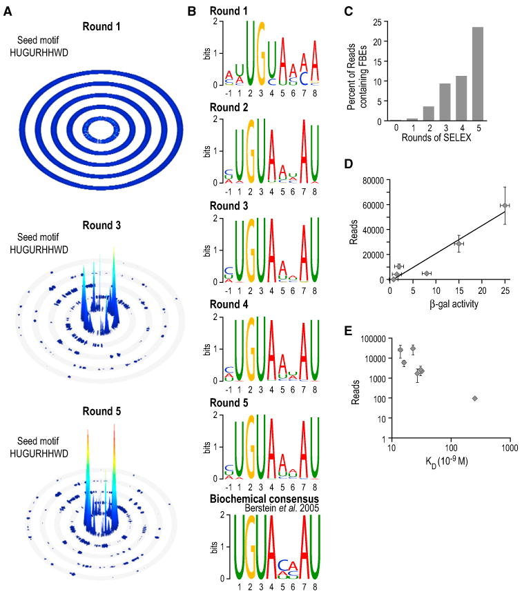
The control and function of RNA are governed by the specificity of RNA binding proteins. Here, we describe a method for global unbiased analysis of RNA-protein interactions that uses in vitro selection, high-throughput sequencing, and sequence-specificity landscapes. The method yields affinities for a vast array of RNAs in a single experiment, including both low- and high-affinity sites. It is reproducible and accurate. Using this approach,we analyzed members of the PUF (Pumilio and FBF) family of eukaryotic mRNA regulators. Our data identify effects of a specific protein partner on PUF-RNA interactions, reveal subsets of target sites not previously detected, and demonstrate that designer PUF proteins can precisely alter specificity. The approach described here is, in principle, broadly applicable for analysis of any molecule that binds RNA, including proteins, nucleic acids, and small molecules.


Figures

Figure 1
Figure 1. The SEQRS Approach
(A) Experimental strategy is illustrated. The DNA library contains a random region of 20 bp (orange) between two 20 bp constant regions (blue). Transcription with T7 RNA polymerase yields a pool of RNAs used as the starting material for selection. A sufficient quantity of RNA to cover all possible 20-mer sequences is incubated with recombinant protein. The protein is immobilized on resin (red circles) to enable capture of the RNA protein complex. After repeated wash steps, the remaining RNA is heat eluted from the protein. The RNA is reverse transcribed (RT) back to DNA using a primer complementary to the constant region denoted as primer B. The single-stranded DNA is amplified using a primer set that reintroduces the T7 promoter (green boxes) upstream of primer A. After the desired number of rounds, aliquots of double-stranded DNA are amplified with primers containing adapters appropriate for Illumina sequencing (pink boxes) and unique 6 bp bar code identifiers (gold boxes). The bar codes differ by at least two bases from all other bar codes to minimize misidentification due to sequencing errors. (B) Overview of computational analysis is shown. After sequencing, the 20-mer random regions are binned according to bar code. All possible k-mer sequences (ten in these experiments) from the random 20-mer are determined for each read. Enrichment over library is calculated by normalizing against the library to correct for differences in coupling efficiency for the random DNA library. Using the n-most abundant reads (n typically = 300), sequence logos are generated. Seed motifs for specificity landscapes are generated from these logos. (C) Visualization of binding specificity. All of the data from an experiment are visualized relative to the seed motif. In this example using C. elegans FBF-2, all of the observed data are fit to the seed motif HUGURWWHD. In the linear form of the inner ring, all possible permutations are arranged in alphabetical order and then the flanking regions are considered. Each ring in the SSL represents increasing numbers of mismatches or hamming distance from the seed motif (shown in blue boxes). The height of each peak is proportional to the enrichment score of a particular sequence. A linearized rendition of the 0-mismatch (innermost) ring is shown at the top of this panel, with sequences indicated.
Figure 2
Figure 2. Enrichment of High-Affinity Sites during Selection
(A) SSLs of FBF-2 at discrete points during selection are shown. The seed motif HUGURHHWD was used for each of the plots. (B) Sequence logos for FBF-2 after five rounds of selection are illustrated. The height of each letter is proportional to the prevalence of that base at the indicated position. A prior selection experiment for RNAs bound by FBF-1 is presented (Bernstein et al., 2005). (C) Enrichment versus rounds of selection is presented. The percentage of sequencing reads containing canonical FBEs defined as UGUNNNAU presented as a function of progression through the cycle. (D) Number of reads versus yeast three-hybrid assays of RNA-protein interactions is shown. LacZ reporter activity in the yeast three-hybrid assay, which is directly correlated with binding affinity, is plotted versus number of reads in SEQRS (Bernstein et al., 2005). Error bars indicate SD. (E) Number of reads versus binding affinity in vitro is illustrated. KD values measured through gel shift assays are compared to the number of reads in SEQRS. Data from analysis of S. cerevisiae Puf4p are shown (Hook et al., 2007; Miller et al., 2008). Error bars indicate SD. The consensus derived from RIP-ChIP is comparable to the motif obtained using SEQRS (Figure S1A).
Figure 3
Figure 3. Analysis of the FBF-2/CPB-1 Protein Complex Reveals Changes in Specificity
(A) Analysis of FBF-2 after five rounds of selection is illustrated. The analysis reveals a motif enriched for an upstream C. The two highest intensity peaks on the SSL both represent sequences with C at the −1 position. (B) Analysis of the CPB-1/FBF-2 complex is presented. The complex yields a distinct motif, as noted in the text. (C) Analysis of the −1 position is shown. The enrichment of −1C is diminished across the entire data set for the CPB-1/FBF-2 complex. (D) A linear representation of the 0-mismatch SSL ring is illustrated. The y axis represents the prevalence of all permutations of the HUGURHHWD motif. Note the lack of enrichment for the upstream C element for the CPB-1/FBF-2 complex. (E) Design of the modified yeast three-hybrid assay is presented. Candidate RNAs were expressed in yeast expressing an FBF-2/AD fusion and the interacting peptide derived from CPB-1. CPB-1 was fused to an SV40 nuclear localization signal, but not to any other domain. Levels of activity of β-galactosidase, produced from the LacZ reporter gene, were used to assay FBF-2 binding to the RNA. (F) CPB-1 enhances binding by FBF-2 to a specific RNA measured by a modified yeast-three hybrid assay. This experiment was done in presence and absence of CPB-1, as indicated below the bars. The gld-1a RNA serves as a positive control for binding. Error bars indicate SD. (G) Additional RNAs assayed using the modified yeast three-hybrid assay are shown. The sequences of additional RNAs analyzed are provided. Data represent the ratio of β-galactosidase levels with and without CPB-1. (H) Design of in vitro translation assays is presented. Repression by FBF-2 was assayed in the presence and absence of CPB-1 in rabbit reticulocyte lysate (RRL). (I) Repression of SEQRS RNA-1 is dependent upon CPB-1. All of the samples were normalized to a mock assay containing only CPB-1. Repression by FBF-2 is insignificant in the absence of CPB-1 or the presence of an interaction-defective version of CPB-1 (FBF-2def). Mutant versions of FBF-2 (Y479A, CPB-1 binding defective, CPBdef, and H326A RNA binding defective, RNAdef) fail to promote repression in the presence of wild-type CPB-1. Error bars indicate SD. SSLs are presented for three additional controls (Figures S2A-S2C).
Figure 4
Figure 4. Specificities of Different PUF Proteins
Sequence logos (above) and SSLs (below) of diverse PUF proteins are illustrated. (A)–(D) present data for a different protein, as indicated. Two seed motifs were used for C. elegans Puf-11 to account for the alternate modes of RNA recognition. Motifs obtained using SEQRS are comparable to those obtained using whole genome approaches for both PUM2 and Puf4p (Figures S1B and S1C).
Figure 5
Figure 5. An Alternate Binding Mode in S. cerevisiae Puf5p
(A) Sequence logos and SSLs are shown. Data were obtained from seven rounds of selection. The combined sequence logo contains two different motifs (denoted “Motif A” and “Motif B”). These differ in the spacing between the UGU and UA elements. (B) Two motifs are presented. SSLs based on motif A (left) and motif B (right) are shown. A well-populated peak containing sequences matching motif A in the 1-mismatch ring of motif B is indicated. Note the difference in the length of separation between the UGU and UA motifs. (C) Design of in vitro repression assay is illustrated. Two luciferase reporter RNAs were combined and incubated in a yeast cell extract. The firefly luciferase reporter contained a putative PUF binding element (PBE); the Renilla reporter did not. The conserved UGU and UA elements are shown in red. Recombinant Puf5p was added to each sample. The ratio of firefly to Renilla luciferase activities was used to quantify effects of the PUF protein on translation (Chritton and Wickens, 2010). The value obtained in a control reaction lacking recombinant Puf5p was used to normalize the data. We tested sites from SPC19 and PRP45 mRNAs because these two mRNAs physically associate with Puf5p, possess motif A in their 3′ UTRs, and lack motif B. Wt, wild-type; Mut, mutant. (D) Puf5p represses translation in vitro via a Motif A (noncanonical) site. Sites from two RNAs physically associated with Puf5p (SPC19 and PRP45) were analyzed (Chritton and Wickens, 2010; Gerber et al., 2004). Both contain Motif A. The Puf5p binding site from CIN8 was used for comparison, and as a positive control. Translation of the firefly luciferase reporter was repressed for all three binding elements. Error bars indicate SD. Motifs A and B are present in 3′ UTRs from transcripts associated with Puf5p in RIP-ChIP assays (Figure S1D).
Figure 6
Figure 6. Specificity of Designer Proteins
(A) Schematic of PUF/RNA complexes and the structure of an FBF-2/RNA complex is presented (Wang et al., 2009b). PUF proteins all possess a similar architecture, in which eight three-helical bundles (green) are stacked into an arc. Along one face of this arc, eight α helices interact with RNA (gray), with a single helix recognizing a single base. Different PUF proteins achieve specificity in part through variations on this basic scaffold. Inset illustrates residues that were altered (N475S and Q479E) and are shown (pink) as sticks opposite the RNA base they coordinate (blue). (B) The sequence logo of the designer protein (N475S Q479E) is shown. The +3 position opposite the site of the N475S and Q479E mutations is indicated with an arrow (pink). Data were obtained after five rounds of selection. (C) Changes in specificity are presented. The relative abundance of sequences fit to the seed motif HUGDRHHWD is shown as a function of position in the 0-mismatch SSL ring in linear form. Sequence families are presented above the x axis. (D and E) The SSL of N475S Q479E after five rounds of selection fit to the wild-type consensus (D) or a motif identified through MEME (E) is illustrated.

References

    1. Ariz M, Mainpal R, Subramaniam K. C. elegans RNA-binding proteins PUF-8 and MEX-3 function redundantly to promote germline stem cell mitosis. Dev Biol. 2009;326:295–304. - PMC - PubMed
    1. Bailey TL, Williams N, Misleh C, Li WW. MEME: discovering and analyzing DNA and protein sequence motifs. Nucleic Acids Res. 2006;34(Web Server issue):W369–W373. - PMC - PubMed
    1. Beerli RR, Barbas CF., 3rd Engineering polydactyl zinc-finger transcription factors. Nat Biotechnol. 2002;20:135–141. - PubMed
    1. Bernstein D, Hook B, Hajarnavis A, Opperman L, Wickens M. Binding specificity and mRNA targets of a C. elegans PUF protein, FBF-1. RNA. 2005;11:447–458. - PMC - PubMed
    1. Besse F, Ephrussi A. Translational control of localized mRNAs: restricting protein synthesis in space and time. Nat Rev Mol Cell Biol. 2008;9:971–980. - PubMed

Publication types

MeSH terms

LinkOut - more resources