Review
. 2012 Sep;13(6):524-46.
doi: 10.2174/138920312803582951.

The eukaryotic flagellum makes the day: novel and unforeseen roles uncovered after post-genomics and proteomics data

Affiliations
Free PMC article
Review

The eukaryotic flagellum makes the day: novel and unforeseen roles uncovered after post-genomics and proteomics data

Michely C Diniz et al. Curr Protein Pept Sci. 2012 Sep.
Free PMC article

Abstract

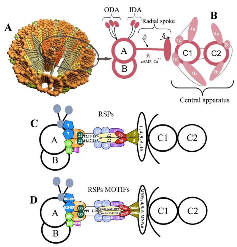
This review will summarize and discuss the current biological understanding of the motile eukaryotic flagellum, as posed out by recent advances enabled by post-genomics and proteomics approaches. The organelle, which is crucial for motility, survival, differentiation, reproduction, division and feeding, among other activities, of many eukaryotes, is a great example of a natural nanomachine assembled mostly by proteins (around 350-650 of them) that have been conserved throughout eukaryotic evolution. Flagellar proteins are discussed in terms of their arrangement on to the axoneme, the canonical "9+2" microtubule pattern, and also motor and sensorial elements that have been detected by recent proteomic analyses in organisms such as Chlamydomonas reinhardtii, sea urchin, and trypanosomatids. Such findings can be remarkably matched up to important discoveries in vertebrate and mammalian types as diverse as sperm cells, ciliated kidney epithelia, respiratory and oviductal cilia, and neuro-epithelia, among others. Here we will focus on some exciting work regarding eukaryotic flagellar proteins, particularly using the flagellar proteome of C. reinhardtii as a reference map for exploring motility in function, dysfunction and pathogenic flagellates. The reference map for the eukaryotic flagellar proteome consists of 652 proteins that include known structural and intraflagellar transport (IFT) proteins, less wellcharacterized signal transduction proteins and flagellar associated proteins (FAPs), besides almost two hundred unannotated conserved proteins, which lately have been the subject of intense investigation and of our present examination.


Figures

Fig. (1)
Fig. (1)
(A) Atomic force microscopy (AFM) contact mode image of an isolated promastigote form of Leishmania chagasi showing its elongated cell body with a single flagellum in surface topography. (B) Contact mode 3D view.
Fig. (2)
Fig. (2)
Schematic illustration of axonemal sub-structures of the eukaryotic flagellum. Panel A, an overview of its internal, axonemal architecture showing the 9+2 cannonical pattern of two inner, central pair (CP) microbules and nine outer microtubule doublets (in golden color), each named A-tubule and B-tubule. Note the presence of radial spokes (RS) and RS proteins (RSPs) as T-shaped structures (in gray) extending from the A-tubule of each doublet to the center of the axoneme (CP), as depicted in Panel B, illustrating the interconnected elements of RSPs, outer dynein arms (ODAs) in green and inner dynein arms (IDAs) in yellow, CP projections in orange. Panel C is the hypothetical “stalk” & “head” model of RSP as a mechanochemical transducer extending from the 9+2 and anchored near the base of the IDA, where the signal input includes calcium binding and/or mechanical strain induced by transient interaction of the spoke head with the central apparatus. Adapted from (and modified after) [113]. Since CaM anchored to the axoneme is a key calcium sensor, while central apparatus and RS are integral elements of calcium signaling pathway [218], four different CaM-interacting protein complexes have been localized: i) to the stalk, RSP2; ii) to the base of the spoke, FAP91; iii and iv) to CP projections, FAP101 and FAP221 [113], with three of these homologs being present in Leishmania genomes (CAC14327, CAB71185 and LmjF35.0290). Several models, including those proposed by [113], [148] and [218], are considered in Panel D, which stands for the probable locations of the RSPs, and in Panel E, which stands for their molecular modules relative to a CP microtubule (right) and an IDA on an outer doublet (left).
Fig. (3)
Fig. (3)
Three-dimensional structures of the first intraflagellar transport (IFT) proteins deposited at Protein Data Bank (PDB). The Chlamydomonas reinhardtii IFT complex 25/27 can be seen on panels A (PDB ID 2CY2) and B (2CY4) [110]. Images are viewed after PDB access modifications made in RCSB PDB Protein Worshop 3.9®.
Fig. (4)
Fig. (4)
Three-dimensional structure of cofilin. A) A 3D model of Leishmania infantum cofilin after B) the PDB template 1QVP_A. A significantly well conserved display of secondary and tertiary structural features can be seen and easily correlated to the average 37% overall similarity between the two primary sequences. Both cofilins have a central mixed β-sheet, which is sandwiched between two pairs of α- helices. The highly conserved residues said to be important for protein stability and correct folding (Tyr64, Trp88, Pro90, and Tyr101, with the exception of Phe85) are present in all Leishmania cofilin sequences and shown in L. infantum modeled cofilin.

Similar articles

Cited by

References

    1. Lewin RA. Studies on the Flagella of Algae. II. Formation of Flagella by Chlamydomonas in Light and Darkness. Annals New York Acad. Sciences. 1953;56:1091–1093. - PubMed
    1. Lewin RA. Mutants of Chlamydomonas moewusii with impaired motility. J. Gen. Microbiol. 1954;11:358–363. - PubMed
    1. Gibbons I R, Grimstone AV. On Flagellar Structure of Certain Flagellates. J. Biophys. Biochem. Cytol. 1960;7:697–716. - PMC - PubMed
    1. Gibbons IR, Rowe AJ. Dynein: a protein with adenosine triphosphatase activity from cilia. Science. 1965;149:424–426. - PubMed
    1. Ringo DL. Flagellar motion and fine structure of the flagellar apparatus in Chlamydomonas. J. Cell Biol. 1967;33:543–571. - PMC - PubMed

Publication types

Substances

LinkOut - more resources