

Hyperintense putaminal rim at 1.5 T: prevalence in normal subjects and distinguishing features from multiple system atrophy
- PMID: 22708511
- PMCID: PMC3460737
- DOI: 10.1186/1471-2377-12-39
Hyperintense putaminal rim at 1.5 T: prevalence in normal subjects and distinguishing features from multiple system atrophy
Abstract
Background: Hyperintense putaminal rim (HPR) is an important magnetic resonance imaging (MRI) sign for multiple system atrophy (MSA). Recent studies have suggested that it can also be observed in normal subjects at 3 T. Whether it can be observed in normal subjects at 1.5 T is not known. This study aimed to determine whether HPR could be observed in normal subjects at 1.5 T; and if so, to establish its prevalence, the MRI characteristics, and the features which distinguish from HPR in MSA patients.
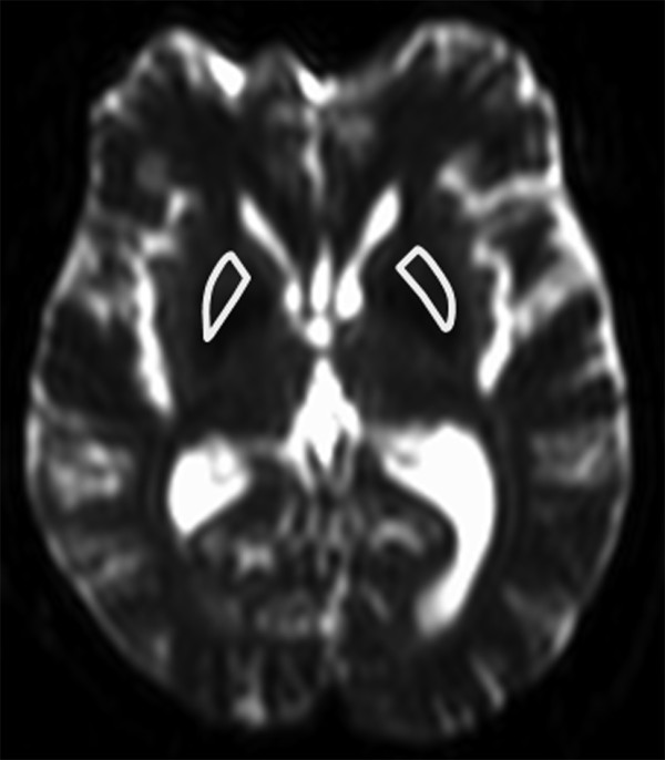
Methods: Axial T2-weighted images of 130 normal subjects were evaluated for the prevalence of HPR, its age and gender distribution, laterality, maximum dimension, association with hypointensity of nearby putamen, and presence of discontinuity. To distinguish from that observed in MSA, axial T2-weighted images of 6 MSA patients with predominant parkinsonism (MSA-P) and 15 MSA patients with predominant cerebellar symptoms (MSA-C) were also evaluated. The characteristics of HPR were compared between these patients and age-matched normal subjects. The mean diffusivity (MD) values of putamen were also compared. Fisher's exact test, t-test, and one way analysis of variance were used to determine significance at corrected p < 0.05.
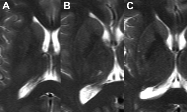
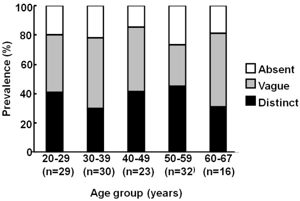
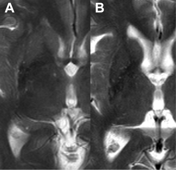
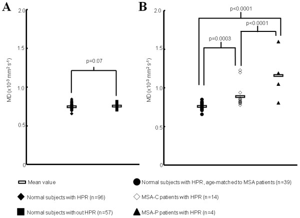
Results: HPR was observed in 38.5% of normal subjects. Age and gender predilection and laterality were not observed. In most cases, it occupied the full length or anterior half of the lateral margin of putamen, and was continuous throughout its length. Maximum transverse dimension was 2 mm. There was no association with hypointensity of nearby putamen. However, in MSA-P, HPR was located predominantly at the posterolateral aspect of putamen, and associated with putaminal atrophy. Discontinuity of HPR was more frequently observed in MSA-P. On visual analysis, the characteristics of HPR were similar between MSA-C patients and normal subjects. Patients with MSA of either type had significantly higher MD values of putamen than normal subjects.
Conclusions: HPR can be observed in 38.5% of normal subjects at 1.5 T. Thin linear hyperintensity without discontinuity, occupying the full length or anterior half of the lateral margin of the putamen, is suggestive of "normal." In doubtful cases, measurement of the MD values of nearby putamen may be valuable.
Figures
References
-
- Savoiardo M, Strada L, Girotti F, Zimmerman RA, Grisoli M, Testa D, Petrillo R. Olivopontocerebellar atrophy: MR diagnosis and relationship to multisystem atrophy. Radiology. 1990;174(3):693–696. - PubMed
Publication types
MeSH terms
LinkOut - more resources
Full Text Sources
Medical
