

Treatment of diabetic mice with undenatured whey protein accelerates the wound healing process by enhancing the expression of MIP-1α, MIP-2, KC, CX3CL1 and TGF-β in wounded tissue
- PMID: 22708778
- PMCID: PMC3676145
- DOI: 10.1186/1471-2172-13-32
Treatment of diabetic mice with undenatured whey protein accelerates the wound healing process by enhancing the expression of MIP-1α, MIP-2, KC, CX3CL1 and TGF-β in wounded tissue
Abstract
Background: Continuous diabetes-associated complications are a major source of immune system exhaustion and an increased incidence of infection. Diabetes can cause poor circulation in the feet, increasing the likelihood of ulcers forming when the skin is damaged and slowing the healing of the ulcers. Whey proteins (WPs) enhance immunity during childhood and have a protective effect on some immune disorders. Therefore, in this study, we investigated the effects of camel WP on the healing and closure of diabetic wounds in a streptozotocin (STZ)-induced type I diabetic mouse model.
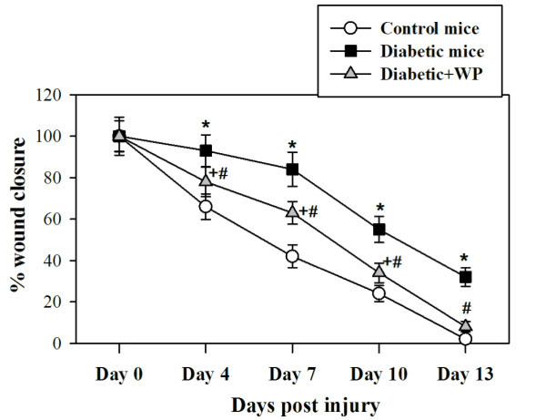
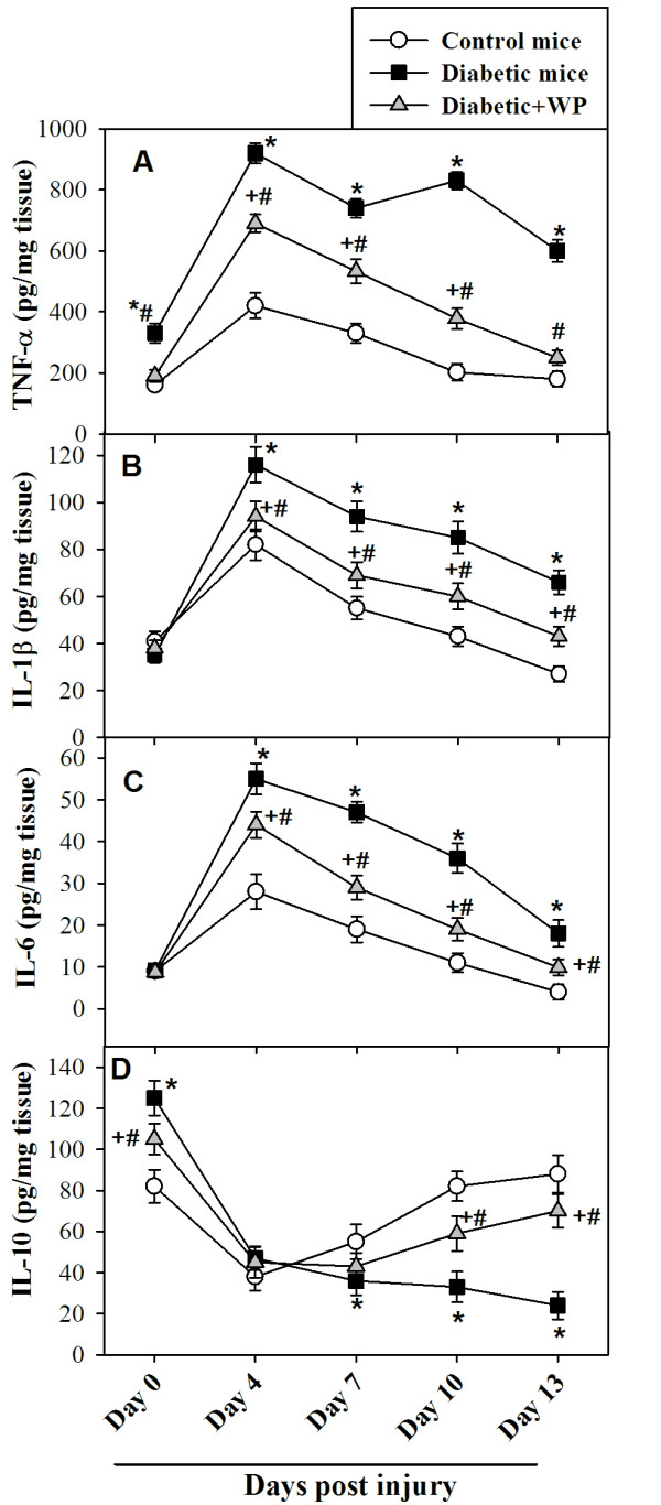
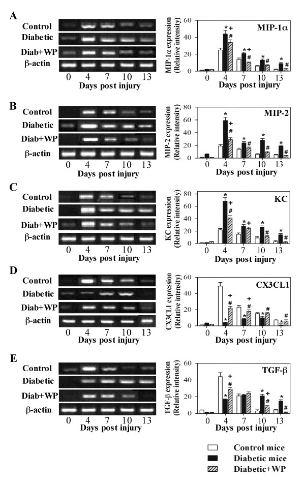
Results: Diabetic mice exhibited delayed wound closure characterized by a significant decrease in an anti-inflammatory cytokine (namely, IL-10) and a prolonged elevation of the levels of inflammatory cytokines (TNF-α, IL-1β and IL-6) in wound tissue. Moreover, aberrant expression of chemokines that regulate wound healing (MIP-1α, MIP-2, KC and CX3CL1) and growth factors (TGF-β) were observed in the wound tissue of diabetic mice compared with control nondiabetic mice. Interestingly, compared with untreated diabetic mice, supplementation with WP significantly accelerated the closure of diabetic wounds by limiting inflammatory stimuli via the restoration of normal IL-10, TNF-α, IL-1β and IL-6 levels. Most importantly, the supplementation of diabetic mice with WP significantly modulated the expression of MIP-1α, MIP-2, KC, CX3CL1 and TGF-β in wound tissue compared with untreated diabetic mice.
Conclusion: Our data demonstrate the benefits of WP supplementation for improving the healing and closure of diabetic wounds and restoring the immune response in diabetic mice.
Figures
Similar articles
-
Camel whey protein enhances diabetic wound healing in a streptozotocin-induced diabetic mouse model: the critical role of β-Defensin-1, -2 and -3.Lipids Health Dis. 2013 Apr 1;12:46. doi: 10.1186/1476-511X-12-46. Lipids Health Dis. 2013. PMID: 23547923 Free PMC article.
-
Supplementation with undenatured whey protein during diabetes mellitus improves the healing and closure of diabetic wounds through the rescue of functional long-lived wound macrophages.Cell Physiol Biochem. 2012;29(3-4):571-82. doi: 10.1159/000338511. Epub 2012 Apr 3. Cell Physiol Biochem. 2012. PMID: 22508064
-
Limiting prolonged inflammation during proliferation and remodeling phases of wound healing in streptozotocin-induced diabetic rats supplemented with camel undenatured whey protein.BMC Immunol. 2013 Jul 25;14:31. doi: 10.1186/1471-2172-14-31. BMC Immunol. 2013. PMID: 23883360 Free PMC article.
-
Wound fluid sampling methods and analysis of cytokine mRNA expression in ulcers from patients with diabetes mellitus.Regen Ther. 2024 Jul 6;26:425-431. doi: 10.1016/j.reth.2024.06.016. eCollection 2024 Jun. Regen Ther. 2024. PMID: 39045578 Free PMC article. Review.
-
Immunomodulatory biomaterials on chemokine signaling in wound healing.Front Pharmacol. 2023 Apr 21;14:1084948. doi: 10.3389/fphar.2023.1084948. eCollection 2023. Front Pharmacol. 2023. PMID: 37153787 Free PMC article. Review.
Cited by
-
Limited Treatment Options for Diabetic Wounds: Barriers to Clinical Translation Despite Therapeutic Success in Murine Models.Adv Wound Care (New Rochelle). 2021 Aug;10(8):436-460. doi: 10.1089/wound.2020.1254. Epub 2020 Dec 18. Adv Wound Care (New Rochelle). 2021. PMID: 33050829 Free PMC article.
-
Exploiting human CD34+ stem cell-conditioned medium for tissue repair.Mol Ther. 2014 Jan;22(1):149-59. doi: 10.1038/mt.2013.194. Epub 2013 Aug 28. Mol Ther. 2014. PMID: 23985698 Free PMC article.
-
Bee gomogenat enhances the healing process of diabetic wounds by orchestrating the connexin-pannexin gap junction proteins in streptozotocin-induced diabetic mice.Sci Rep. 2023 Nov 15;13(1):19961. doi: 10.1038/s41598-023-47206-5. Sci Rep. 2023. PMID: 37968314 Free PMC article.
-
Collagen-Containing Fish Sidestream-Derived Protein Hydrolysates Support Skin Repair via Chemokine Induction.Mar Drugs. 2021 Jul 15;19(7):396. doi: 10.3390/md19070396. Mar Drugs. 2021. PMID: 34356821 Free PMC article.
-
Why whey? Camel whey protein as a new dietary approach to the management of free radicals and for the treatment of different health disorders.Iran J Basic Med Sci. 2017 Apr;20(4):338-349. doi: 10.22038/IJBMS.2017.8573. Iran J Basic Med Sci. 2017. PMID: 28804604 Free PMC article. Review.
References
-
- Lioupis C. Effects of diabetes mellitus on wound healing: an update. J Wound Care. 2005;14:84–86. - PubMed
-
- Nithya V, Baskar A. A Preclinical Study on Wound Healing Activity of Lawsonia ulba Linn. Res J of Phytochemistry. 2011;5:123–129.
Publication types
MeSH terms
Substances
LinkOut - more resources
Full Text Sources
Medical
Research Materials
Miscellaneous
