

Review
doi: 10.1111/j.1365-2818.2012.03619.x.
Epub 2012 Jun 18.
Optical coherence tomography
Affiliations
- PMID: 22708800
- PMCID: PMC3563006
- DOI: 10.1111/j.1365-2818.2012.03619.x
Item in Clipboard
Review
Optical coherence tomography
J Microsc.
2012 Sep.
Free PMC article
Abstract
The review provides a concise explanation of principles of operation of different optical coherence tomography methods. A comparative analysis of their advantages and disadvantages is presented in relation to specific applications. The review will assist the reader in making an educated choice on the most suitable optical coherence tomography method to be used in a particular application.
© 2012 The Authors Journal of Microscopy © 2012 Royal Microscopical Society.
Figures
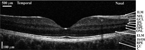
Cross-section image of the retina using an en face time domain OCT system driven by a large bandwidth source (Cucu et al., 2006). Lateral size: 7.5 mm, vertical size is along depth, 0.725 mm measured in the retina. ILM, inner limiting membrane; NFL, nerve fibre layer; GCL, ganglion cell layer; IPL, inner plexiform layer; INL, inner nuclear layer; OPL, outer plexiform layer; ONL, outer nuclear layer; ELM, external limiting membrane; IS/OS, junction between the inner and outer photoreceptors; RPE, retinal pigment epithelium; CC, choriocapillaris; C, choroid.
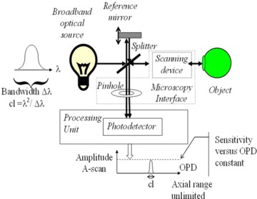
TD-OCT setup.
TD-OCT: Relative size of the coherence length, cl, required depth resolution δz, and OPD axial range 2Δz determined by the axial extension of the Object to be investigated. For simplicity, the index of refraction is considered as 1.
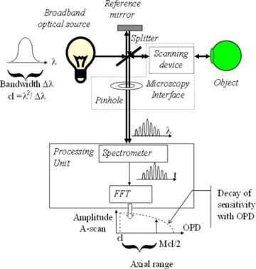
SB-OCT setup.
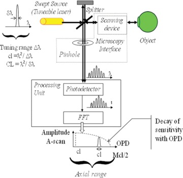
SS-OCT setup.
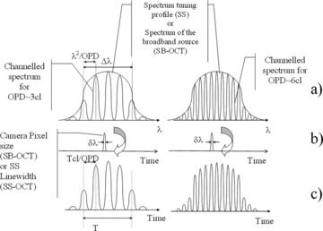
(a) Spectrum output of the interferometer for two OPD values, left: ∼3 cl, right: ∼6 cl; (b) SD-OCT sampling function, of width δλ, spectrum is read in a time T either by downloading the charge from the linear camera array, or by tuning the frequency of the SS, the large arrow suggests that by doing so, the spectrum shown above in (a) is transferred to the output signal shown in (c) below; (c) signal delivered by the Processing unit in Figures 4 and 5.

SD-OCT: Relative size of the CL extension of interfering wavetrains, required depth resolution δz, and axial range Δz determined by the axial extension of the Object to be investigated.
References
-
- Bachmann AH, Leitgeb RA, Lasser T. Heterodyne Fourier domain optical coherence tomography for full range probing with high axial resolution. Opt. Express. 2006;14(4):1487–1496. - PubMed
-
- Bonin T, Franke G, Hagen-Eggert M, Koch P, Hüttmann G. In vivo fourier-domain full-field oct of the human retina with 1.5 million a-lines/s. Opt. Lett. 2010;35(20):3432–3434. - PubMed
-
- Bradu A, Podoleanu AGh. Attenuation of mirror image and enhancement of the signal-to-noise ratio in a Talbot bands optical coherence tomography system. J. Biomed. Opt. 2011;16(7):076010-1–10. - PubMed
-
- Cense B, Chen TC, Park BH, Pierce MC, de Boer JF. In vivo birefringence and thickness measurements of the human retinal nerve fibre layer using polarization-sensitive optical coherence tomography. J Biomed Opt. 2004;9(1):121–125. - PubMed
-
- Cucu RG, Podoleanu AGh, Rogers JA, Pedro J, Rosen RB. Combined confocal scanning ophthalmoscopy/en face T-scan based ultrahigh resolution OCT of the human retina in vivo. Opt. Lett. 2006;31(11):1684–1687. - PubMed
Publication types
MeSH terms
Grants and funding
LinkOut - more resources
Full Text Sources
Other Literature Sources
Medical
