. 2012 Jun 18:12:13.
doi: 10.1186/1472-6807-12-13.

Crystal structure of the Yersinia enterocolitica type III secretion chaperone SycD in complex with a peptide of the minor translocator YopD

Affiliations

Crystal structure of the Yersinia enterocolitica type III secretion chaperone SycD in complex with a peptide of the minor translocator YopD

Madeleine Schreiner et al. BMC Struct Biol. .

Abstract

Background: Type III secretion systems are used by Gram-negative bacteria as "macromolecular syringes" to inject effector proteins into eukaryotic cells. Two hydrophobic proteins called translocators form the necessary pore in the host cell membrane. Both translocators depend on binding to a single chaperone in the bacterial cytoplasm to ensure their stability and efficient transport through the secretion needle. It was suggested that the conserved chaperones bind the more divergent translocators via a hexapeptide motif that is found in both translocators and conserved between species.

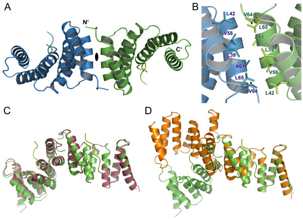
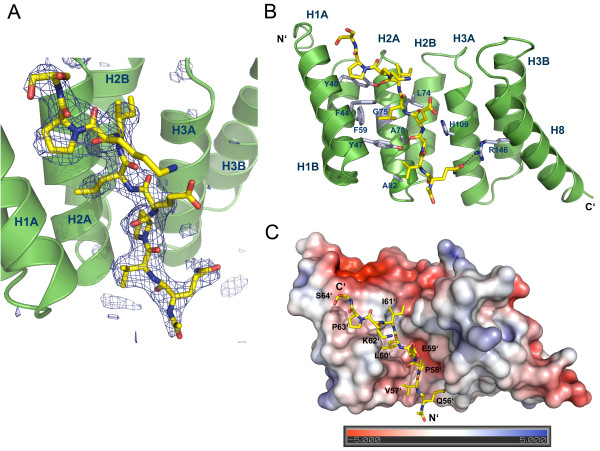
Results: We crystallized a synthetic decapeptide from the Yersinia enterocolitica minor type III secretion translocator YopD bound to its cognate chaperone SycD and determined the complex structure at 2.5 Å resolution. The structure of peptide-bound SycD is almost identical to that of apo SycD with an all helical fold consisting of three tetratricopeptide repeats (TPRs) and an additional C-terminal helix. Peptide-bound SycD formed a kinked head-to-head dimer that had previously been observed for the apo form of SycD. The homodimer interface comprises both helices of the first tetratricopeptide repeat. The YopD peptide bound in extended conformation into a mainly hydrophobic groove on the concave side of SycD. TPRs 1 and 2 of SycD form three hydrophobic pockets that accommodated the conserved hydrophobic residues at position 1, 3 and 6 of the translocator hexapeptide sequence. Two tyrosines that are highly conserved among translocator chaperones contribute to the hydrophobic patches but also form hydrogen bonds to the peptide backbone.

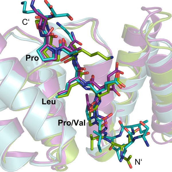
Conclusions: The interaction between SycD and YopD is very similar to the binding of the Pseudomonas minor translocator PopD to its chaperone PcrH and the Shigella major translocator IpaB to its chaperone IpgC. This confirms the prediction made by Kolbe and co-workers that a hexapeptide with hydrophobic residues at three positions is a conserved chaperone binding motif. Because the hydrophobic groove on the concave side of translocator chaperones is involved in binding of the major and the minor translocator, simultaneous binding of both translocators to a single type III secretion class II chaperone appears unlikely.


Figures

Figure 1
Figure 1
Dimer arrangement of the SycD:YopD complex. (A) Peptide-bound SycD forms a symmetric dimer, in which protomers are related by a crystallographic 2-fold axis. The dimerization is mediated via the two N-terminal helices (TPR1). (B) Dimerization interface of the SycD:YopD complex stabilized by van der Waal contacts involving the labeled residues shown as sticks. (C) Pairwise alignment (DaliLite server [29]) of SycD:YopD (green) to kinked apo SycD (red) (PDB ID: 2VGY) and to (D) monomer B of elongated apo SycD (orange) (PDB ID: 2VGX) shows that the dimer arrangement of the chaperone-peptide complex is nearly identical to the arrangement of the kinked apo form.
Figure 2
Figure 2
Structure of SycD in complex with the YopD peptide. (A) Positive difference electron density within the Fo-Fc map contoured at 3σ (blue mesh) assigns the position of the YopD peptide (yellow) to the concave groove of SycD (green). The Fo-Fc map was calculated via simulated annealing refinement in PHENIX [30] using the fully refined structural model whose YopD peptide chain was removed prior to refinement. (B) The YopD peptide lies antiparallel to helix H1A within the concave cleft interacting with various mainly hydrophobic residues lining H1A, H2A and H3A. Hydrogen bonds (purple lines) involving Tyr40, Tyr47 and Arg146 additionally stabilize the complex. (C) Electrostatic surface potential of the SycD:YopD complex. Three nonpolar residues Pro58’, Pro63’ and Leu60’ occupy distinct hydrophobic pockets and anchor the peptide (yellow) into the concave cleft. Negatively charged surface areas are colored in red, positively charged areas in blue.
Figure 3
Figure 3
Superposition of the chaperone binding domains of T3S translocators. The superposition (DaliLite server [29]) of the chaperone-peptide complexes SycD:YopD (green), PcrH:PopD (purple) (PDB ID: 2XCB, chain A) and IpgC:IpaB (cyan) (PDB ID: 3GZ1, chain B) reveals a good spatial agreement within the key anchor residues of the YopD, PopD and IpaB sequence motifs.

Similar articles

Cited by

References

    1. Dean P. Functional domains and motifs of bacterial type III effector proteins and their roles in infection. FEMS Microbiol Rev. 2011;35:1100–1125. doi: 10.1111/j.1574-6976.2011.00271.x. - DOI - PubMed
    1. Trosky JE, Liverman ADB, Orth K. Yersinia outer proteins: Yops. Cell Microbiol. 2008;10:557–565. doi: 10.1111/j.1462-5822.2007.01109.x. - DOI - PubMed
    1. Cornelis GR. The type III secretion injectisome. Nat Rev Micro. 2006;4:811–825. doi: 10.1038/nrmicro1526. - DOI - PubMed
    1. Izoré T, Job V, Dessen A. Biogenesis, Regulation, and Targeting of the Type III Secretion System. Structure. 2011;19:603–612. doi: 10.1016/j.str.2011.03.015. - DOI - PubMed
    1. Troisfontaines P, Cornelis GR. Type III Secretion: More Systems Than You Think. Physiology. 2005;20:326–339. doi: 10.1152/physiol.00011.2005. - DOI - PubMed

Publication types

MeSH terms

LinkOut - more resources