. 2012 Oct;7(10):1507-19.
doi: 10.2217/nnm.12.27. Epub 2012 Jun 18.

Antiangiogenic nanotherapy with lipase-labile Sn-2 fumagillin prodrug

Affiliations

Antiangiogenic nanotherapy with lipase-labile Sn-2 fumagillin prodrug

Dipanjan Pan et al. Nanomedicine (Lond). 2012 Oct.

Abstract

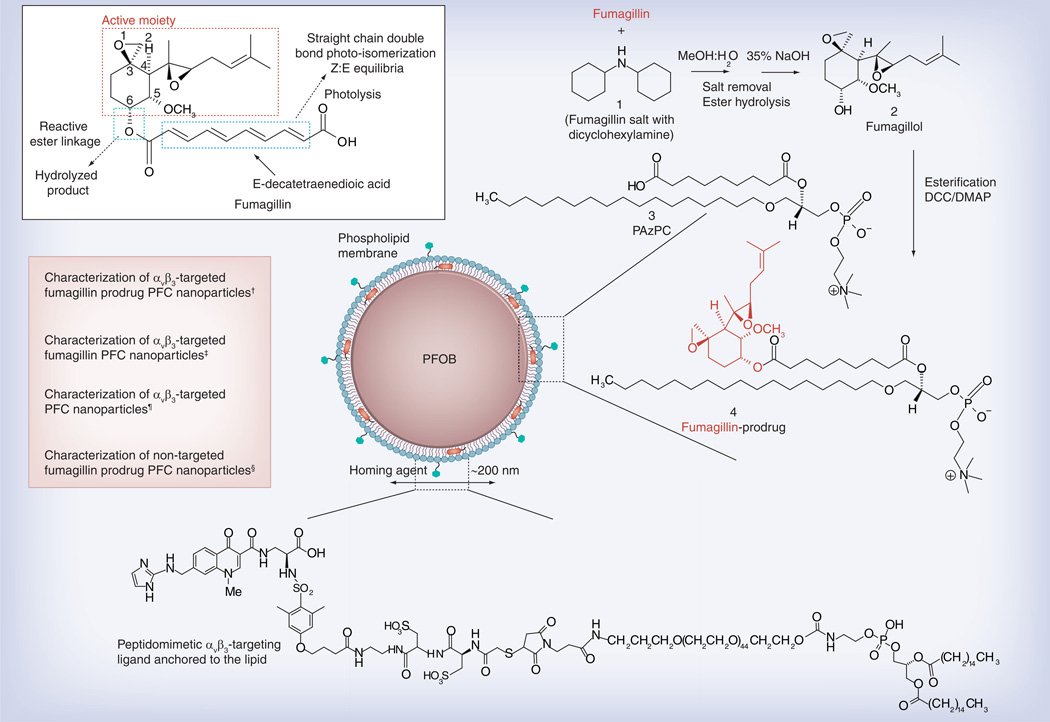
Background: The chemical instability of antiangiogenic fumagillin, combined with its poor retention during intravascular transit, requires an innovative solution for clinical translation. We hypothesized that an Sn-2 lipase-labile fumagillin prodrug, in combination with a contact-facilitated drug delivery mechanism, could be used to address these problems.

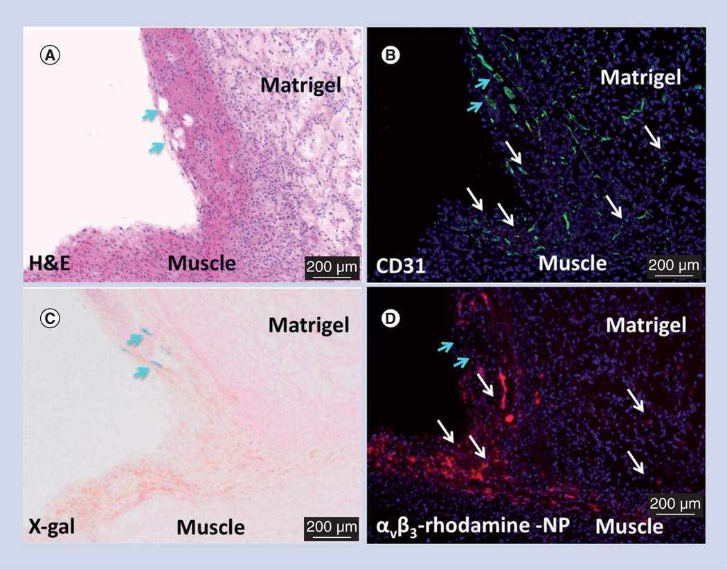
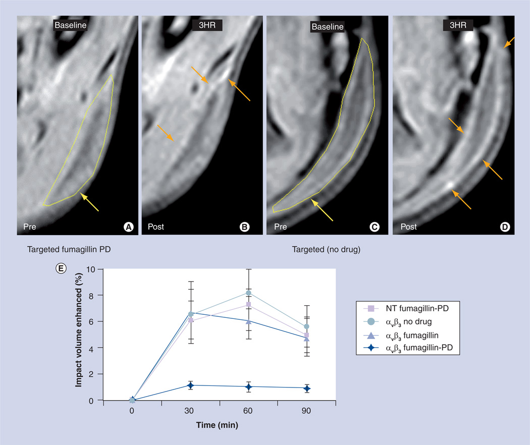
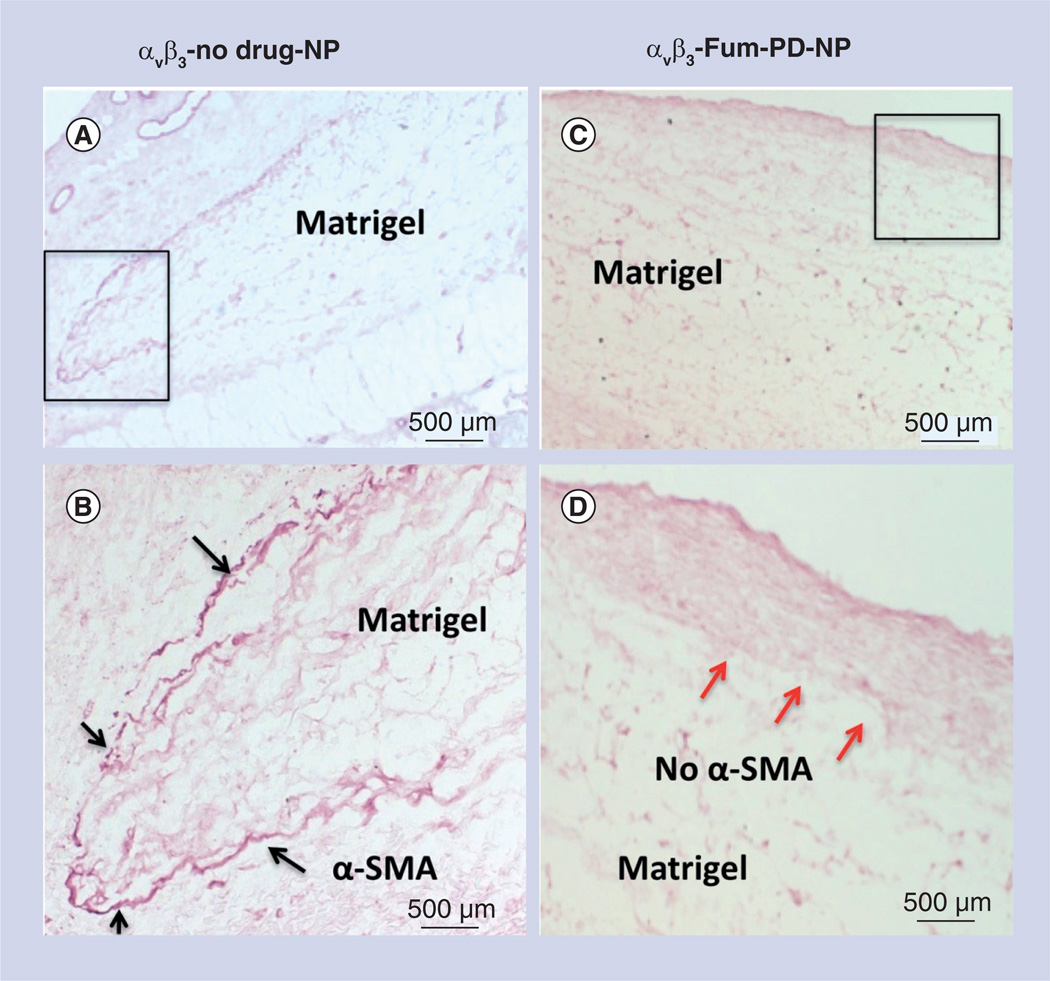
Methods: α(v)β(3)-targeted and nontargeted nanoparticles with and without fumagillin in the prodrug or native forms were evaluated in vitro and in vivo in the Matrigel™ (BD Biosciences, CA, USA) plug model of angiogenesis in mice.

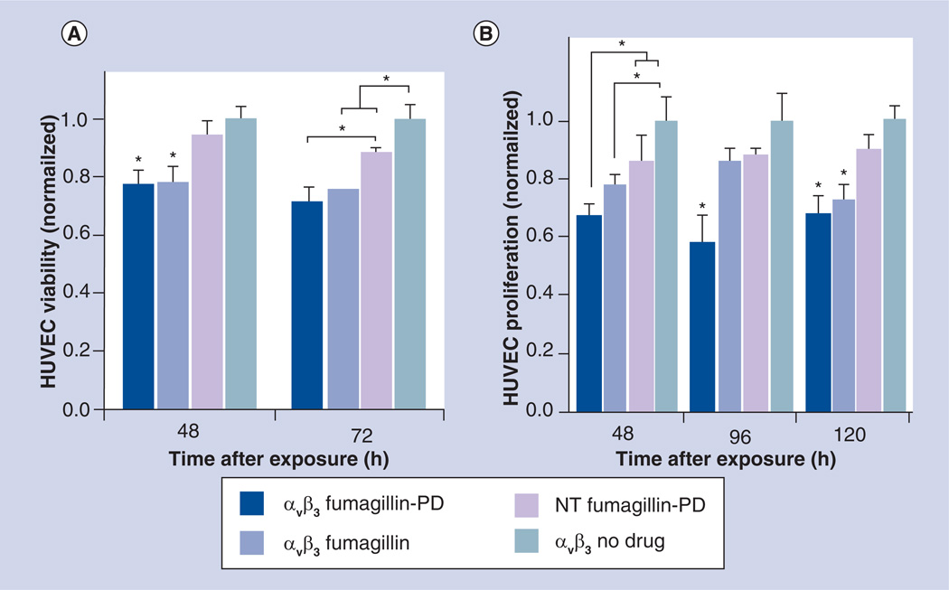
Results: In vitro experiments demonstrated that the new fumagillin prodrug decreased viability at least as efficacious as the parent compound, on an equimolar basis. In the Matrigel mouse angiogenesis model, α(v)β(3)-fumagillin prodrug decreased angiogenesis as measured by MRI (3T), while the neovasculature was unaffected with the control nanoparticles.

Conclusion: The present approach resolved the previously intractable problems of drug instability and premature release in transit to target sites.


Conflict of interest statement

Financial & competing interests disclosure

The authors have no other relevant affiliations or financial involvement with any organization or entity with a financial interest in or financial conflict with the subject matter or materials discussed in the manuscript apart from those disclosed.

No writing assistance was utilized in the production of this manuscript.

Figures

Figure 1
Figure 1. Structure and active sites of fumagillin with their sources of instability indicated (see figure on previous page)
Synthetic strategy for the preparation of Sn-2 fumagillin prodrug and development of site-targeted nanoparticles: saponification of fumagillin with MeOH:water (1:1), 35% NaOH; esterification with PAzPC, DCC/DMAP; preparation of a lipid-thin film from a phospholipid mixture of 98.7 mole% lecithin phosphatidylcholine, 0.15 mole% of αvβ3-ligand-conjugated lipid and 1.12 mole% of fumagillin prodrug; self-assembly by brief sonication and microfluidization, perfluorocarbon, glycerin, pH 6.5, at 20,000 psi for 4 min. Hydrodynamic diameter (dynamic light scattering [DLS]): 220 nm; electrophoretic potential: −17 mV; polydispersity index: 0.19. Hydrodynamic diameter (DLS): 280 nm; electrophoretic potential: −22 mV; polydispersity index: 0.25. Hydrodynamic diameter (DLS): 230 nm; electrophoretic potential: −21 mV; polydispersity index: 0.06. §Hydrodynamic diameter (DLS): 280 nm; electrophoretic potential: −10 mV; polydispersity index: 0.08.
Figure 2
Figure 2. In vitro serial effect of Sn-2 lipase-labile fumagillin prodrug-incorporated nanoparticles on HUVEC cell metabolic activity (A) and proliferation (B)
The αvβ3-targeted Sn-2 phospholipase-labile PD nanoparticles had equivalent effectiveness in vitro on HUVEC (A) cell metabolic activity and (B) proliferation, compared with the αvβ3-targeted native fumagillin nanoparticles used in several previous studies [–6]. Collectively, these data affirm the in vitro sensitivity of proliferating endothelial cells to the effects of αvβ3-targeted fumagillin nanoparticles, included in the native or PD form. *p < 0.05 (indicates significant difference). HUVEC: Human umbilical vein endothelial cell; NT: Nontargeted; PD: Prodrug.
Figure 3
Figure 3. Microscopic examination of FGF Matrigel™ subcutaneous explant (serial sections) from FVB/N-TgN(TIE-2-LacZ)182-Sato mice following injection (intravenous) of αvβ3-targeted rhodamine-labeled perfluorocarbon nanoparticles
(A) H&E staining to illustrate the anatomic location of the implant relative to the muscle, revealing a region with prominent mature vessels (arrows). Note proliferation of inflammatory cells along the muscle and implant interface. (B) PECAM (CD31) staining of endothelium revealing prominent vasculature along the Matrigel periphery and the development of microvessels within the implant. (C) LacZ signal for β-galactosidase under Tie-2 promoter control. Lac-Z staining (arrows) is appreciated in a region of more mature vessels along the implant periphery but the signal was not observed in most of the inflammatory regions along the muscle–implant interface or within the implant. (D) Binding of the αvβ3-targeted rhodamine-labeled perfluorocarbon nanoparticles densely within the inflammed tissue along the muscle and implant interface and diffusely within the Matrigel implant. Regions of αvβ3-targeted rhodamine-labeled perfluorocarbon nanoparticle binding is generally correlated with areas of PECAM staining (white arrows) except for an area of rhodamine signal dropout (blue arrows) in (D), which spatially paralleled prominent PECAM and LacZ staining in (B) and (C), respectively. These images corroborate the specific binding of αvβ3-targeted nanoparticles to nascent angiogenic vessels (Tie-2, PECAM+) and not to maturing microvessels (Tie-2+, PECAM+). H&E: Hematoxylin and eosin; NP: Nanoparticle.
Figure 4
Figure 4. In vivo MRI study of pre- and post-treatment with targeted fumagillin prodrug nanoparticles
In vivo MR signal enhancement post-treatment with (A & B) targeted fumagillin nanoparticles and (C & D) control (no drug). In the pre-treatment images, areas of Matrigel™ implantation have been marked with yellow arrows. In the post-treatment images, signal enhancement has been denoted with orange arrows. (E) Reduced Matrigel implant enhancement in rats treated with αvβ3-integrin-targeted nanoparticles with fumagillin-PD versus αvβ3-integrin-targeted nanoparticles with fumagillin, αvβ3-integrin-targeted nanoparticles without drug and nontargeted nanoparticles with fumagillin-PD. NT: Nontargeted; PD: Prodrug.
Figure 5
Figure 5. Microscopic examination of FGF Matrigel™ subcutaneous explant in mice following serial injection (intravenous) of αvβ3-targeted no-drug perfluorocarbon nanoparticles (αvβ3-no drug-NP) or αvβ3-targeted fumagillin prodrug perfluorocarbon nanoparticles (αvβ3-Fum-PD-NP)
(A) Low power image of Matrigel™ explant from control animal that was immunostained to identify maturing vessels with α-SMA. Positive staining was appreciated peripherally around the majority of the plug (black box). (B) Enlarged view of boxed region in (A), showing the peripheral α-SMA biomarker. (C) Low power image of Matrigel explant from fumagillin PD-treated animal that was immunostained to identify maturing vessels with α-SMA. Virtually no positive staining was appreciated along the implant periphery, suggesting that the repeated pruning of angiogenic vessels with fumagillin PD impaired the development and progression of mature vessels in contrast with the control. (D) Enlarged view of boxed region in (C), illustrating the lack of peripheral α-SMA biomarker (red arrows). α-SMA: α-smooth muscle actin; NP: Nanoparticle; PD: Prodrug.

References

    1. Sin N, Meng L, Wang MQ, Wen JJ, Bornmann WG, Crews CM. The anti-angiogenic agent fumagillin covalently binds and inhibits the methionine aminopeptidase, MetAP-2. Proc. Natl Acad. Sci. USA. 1997;94(12):6099–6103. ▪▪ Introduces the antiangiogenic effect of fumagillin and mechanistic insight into MetAP-2-binding.

    1. Liu S, Widom J, Kemp CW, Crews CM, Clardy J. Structure of human methionine aminopeptidase-2 complexed with fumagillin. Science. 1998;282(5392):1324–1327. ▪ Structural elucidation of fumagillin complexation with human MetAP-2.

    1. Arfin SM, Kendall RL, Hall L et al. Eukaryotic methionyl aminopeptidases: two classes of cobalt-dependent enzymes. Proc. Natl Acad. Sci. USA. 1995;92(17):7714–7718. - PMC - PubMed
    1. Wang J, Lou P, Henkin J. Selective inhibition of endothelial cell proliferation by fumagillin is not due to differential expression of methionine aminopeptidases. J. Cell. Biochem. 2000;77(3):465–473. - PubMed
    1. Lowther WT, Mcmillen DA, Orville AM, Matthews BW. The anti-angiogenic agent fumagillin covalently modifies a conserved active-site histidine in the Escherichia coli methionine aminopeptidase. Proc. Natl Acad. Sci. USA. 1998;95(21):12153–12157. - PMC - PubMed

Patents

    1. Harris TD. M Rajopadhye: US6511648. 2003

Websites

    1. NCI. Developmental Therapeutics Program. www.dtp.nci.nih.gov.
    1. Robinson G. Histological techniques. http://mammary.nih.gov/tools/histological/histology/index.html.

Publication types