

An acidic oligopeptide displayed on AAV2 improves axial muscle tropism after systemic delivery
- PMID: 22709483
- PMCID: PMC3416570
- DOI: 10.1186/1479-0556-10-3
An acidic oligopeptide displayed on AAV2 improves axial muscle tropism after systemic delivery
Abstract
Background: The appropriate tropism of adeno-associated virus (AAV) vectors that are systemically injected is crucial for successful gene therapy when local injection is not practical. Acidic oligopeptides have been shown to enhance drug delivery to bones.
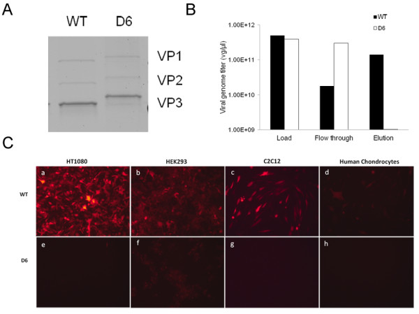
Methods: In this study six-L aspartic acids (D6) were inserted into the AAV2 capsid protein sequence between amino acid residues 587 and 588. 129SVE mice were injected with double-stranded wild-type- (WT-) or D6-AAV2 mCherry expression vectors (3.24 x 10¹⁰ vg per animal) via the superficial temporal vein within 24 hours of birth.
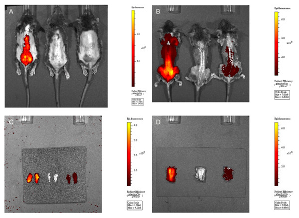
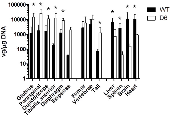
Results: Fluorescence microscopy and quantitative polymerase chain reaction confirmed higher levels of mCherry expression in the paraspinal and gluteus muscles in the D6-AAV2 injected mice. The results revealed that although D6-AAV2 was less efficient in the transduction of immortalized cells stronger mCherry signals were detected over the spine and pelvis by live imaging in the D6-AAV2-injected mice than were detected in the WT-AAV2-injected mice. In addition, D6-AAV2 lost the liver tropism observed for WT-AAV2.
Conclusions: An acidic oligopeptide displayed on AAV2 improves axial muscle tropism and decreases liver tropism after systemic delivery. This modification should be useful in creating AAV vectors that are suitable for gene therapy for diseases involving the proximal muscles.
Figures

Similar articles
-
Focused ultrasound as a novel strategy for noninvasive gene delivery to retinal Müller glia.Theranostics. 2020 Feb 10;10(7):2982-2999. doi: 10.7150/thno.42611. eCollection 2020. Theranostics. 2020. PMID: 32194850 Free PMC article.
-
A muscle-targeting peptide displayed on AAV2 improves muscle tropism on systemic delivery.Gene Ther. 2009 Aug;16(8):953-62. doi: 10.1038/gt.2009.59. Epub 2009 May 28. Gene Ther. 2009. PMID: 19474807 Free PMC article.
-
Anti-EpCAM-conjugated adeno-associated virus serotype 2 for systemic delivery of EGFR shRNA: Its retargeting and antitumor effects on OVCAR3 ovarian cancer in vivo.Acta Biomater. 2019 Jun;91:258-269. doi: 10.1016/j.actbio.2019.04.044. Epub 2019 Apr 23. Acta Biomater. 2019. PMID: 31026519
-
Chimeric Capsid Proteins Impact Transduction Efficiency of Haploid Adeno-Associated Virus Vectors.Viruses. 2019 Dec 9;11(12):1138. doi: 10.3390/v11121138. Viruses. 2019. PMID: 31835440 Free PMC article.
-
Pseudotyped adeno-associated viral vector tropism and transduction efficiencies in murine wound healing.Wound Repair Regen. 2012 Jul-Aug;20(4):592-600. doi: 10.1111/j.1524-475X.2012.00810.x. Epub 2012 Jun 19. Wound Repair Regen. 2012. PMID: 22713157 Free PMC article.
Cited by
-
Tailoring the AAV2 capsid vector for bone-targeting.Pediatr Res. 2018 Oct;84(4):545-551. doi: 10.1038/s41390-018-0095-8. Epub 2018 Oct 15. Pediatr Res. 2018. PMID: 30323349 Free PMC article.
-
Controlling AAV Tropism in the Nervous System with Natural and Engineered Capsids.Methods Mol Biol. 2016;1382:133-49. doi: 10.1007/978-1-4939-3271-9_10. Methods Mol Biol. 2016. PMID: 26611584 Free PMC article.
-
Adeno-Associated Virus Engineering and Load Strategy for Tropism Modification, Immune Evasion and Enhanced Transgene Expression.Int J Nanomedicine. 2024 Jul 29;19:7691-7708. doi: 10.2147/IJN.S459905. eCollection 2024. Int J Nanomedicine. 2024. PMID: 39099791 Free PMC article. Review.
References
LinkOut - more resources
Full Text Sources
Other Literature Sources
