. 2012 Jun 18;12(1):29.
doi: 10.1186/1475-2867-12-29.

Expression of osteoprotegerin, receptor activator of nuclear factor kappa-B ligand, tumor necrosis factor-related apoptosis-inducing ligand, stromal cell-derived factor-1 and their receptors in epithelial metastatic breast cancer cell lines

Affiliations

Expression of osteoprotegerin, receptor activator of nuclear factor kappa-B ligand, tumor necrosis factor-related apoptosis-inducing ligand, stromal cell-derived factor-1 and their receptors in epithelial metastatic breast cancer cell lines

Vivian Labovsky et al. Cancer Cell Int. .

Abstract

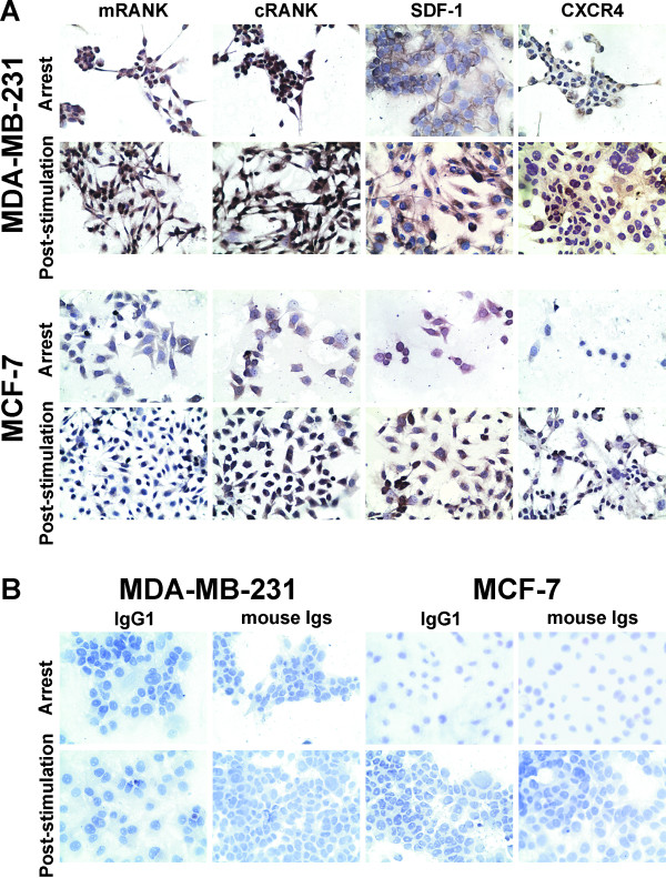
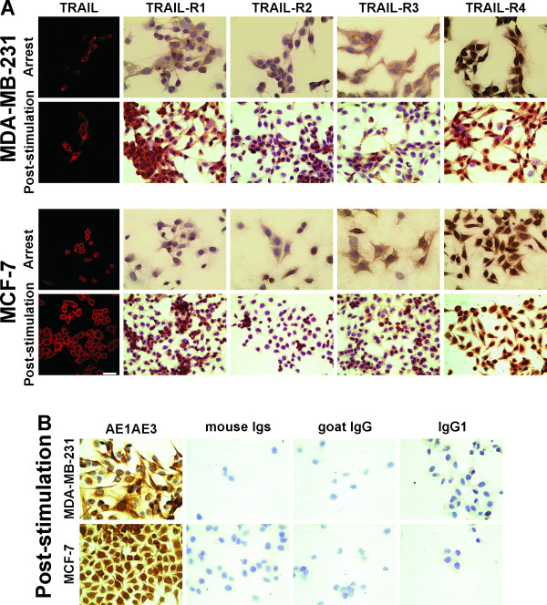
Background: While breast cancer (BC) is the major cause of death among women worldwide, there is no guarantee of better patient survival because many of these patients develop primarily metastases, despite efforts to detect it in its early stages. Bone metastasis is a common complication that occurs in 65-80 % of patients with disseminated disease, but the molecular basis underlying dormancy, dissemination and establishment of metastasis is not understood. Our objective has been to evaluate simultaneously osteoprotegerin (OPG), receptor activator of nuclear factor kappa B ligand (RANKL), tumor necrosis factor-related apoptosis-inducing ligand (TRAIL), stromal cell-derived factor-1 (SDF-1), and their receptors (R) in 2 human BC cell lines, MDA-MB-231 and MCF-7.

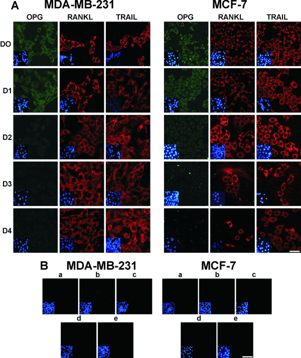
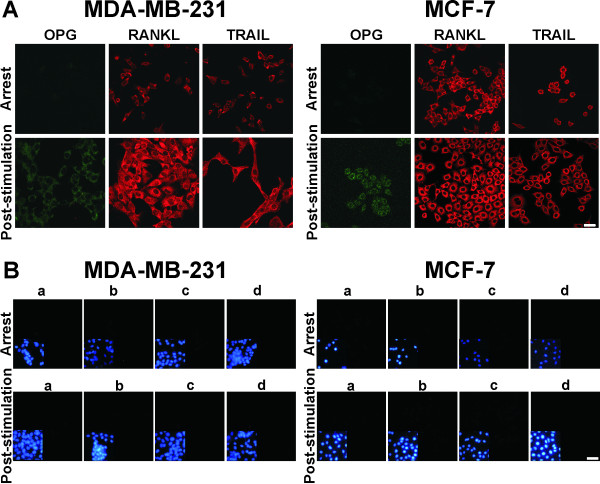
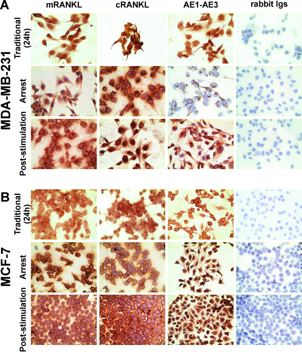
Methods: OPG, RANKL, TRAIL and SDF-1 expression and release, in addition to the expression of their receptors has been investigated using immunofluorescence, immunocytochemistry and ELISA analyses.

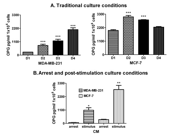
Results: MCF-7 cells released higher levels of OPG in conditioned media (CM) than MDA-MB-231 cells; 100 % of both types of cell expressed OPG, RANKL, TRAIL and SDF-1. Moreover, 100 % in both lines expressed membrane RANKL and RANK, whereas only 50 % expressed CXCR4. Furthermore, 100 % expressed TRAIL-R1 and R4, 30-50 % TRAIL-R2, and 40-55 % TRAIL-R3.

Conclusions: MCF-7 and MDA-MB-231 cells not only released OPG, but expressed RANKL, TRAIL and SDF-1. The majority of the cells also expressed RANK, CXCR4 and TRAIL-R. Since these ligands and their receptors are implicated in the regulation of proliferation, survival, migration and future bone metastasis during breast tumor progression, assessment of these molecules in tumor biopsies of BC patients could be useful in identifying patients with more aggressive tumors that are also at risk of bone metastasis, which may thus improve the available options for therapeutic intervention.


Figures

Figure 1
Figure 1
Release of OPG from BC cell lines under two different culture conditions detected by ELISA. (A) Traditional culture conditions. Conditioned media (CM) from the cell cultures at 24, 48, 72 and 96 h [Days (D): D1-D4] from both BC cell lines were used to evaluate the levels of OPG by ELISA. The values of OPG are expressed as X¯ pg/ml/ 1x10⁶ cells + standard error (SE). Statistical significance: One-way analysis of variance (ANOVA) followed by a Bonferroni post hoc test. Asterisks (*) indicate a significant difference (p <0.05) compared to D1 for each cell line. (B) Arrest and post-stimulation culture conditions. CM obtained under arrest at 48 h and subsequent stimulation with 10 % SBF for 48 h from both BC cell lines were used to evaluate the levels of OPG by ELISA. The OPG values are expressed as X¯ pg/ml/ 1x10⁶ cells + standard error (SE). Statistical significance: Unpaired t-test with Welch's correction. Asterisks (*) indicate a significant difference (p = 0.0158, * and p = 0.0056, **) between OPG production during arrest and after stimulation in the MDA-MB-231 and MCF-7 cell lines.
Figure 2
Figure 2
Immunofluorescence detection of OPG, RANKL and TRAIL in both BC cell lines grown under Traditional culture conditions. (A) Immunofluorescence staining for OPG, RANKL and TRAIL was positive in both BC cell lines. D: day; D0: cells at time zero; D1, D2, D3 and D4: cell lines grown for 24, 48, 72 and 96 h, respectively. The insert shows nuclear DNA stained with DAPI. The scale bar represents 30 μm. (B) Both BC cell lines were grown until D4 (96 h) and incubated either without a primary Ab and with the followings Abs: a) Cy3-labeled goat anti-mouse IgG, b) FITC-labeled goat anti-rabbit IgG, c) an irrelevant Ab as a negative isotype control, such as IgG1, d) mouse Igs and e) rabbit Igs. In controls c and d, the cell lines were stained with a Cy3-labeled goat anti-mouse IgG Ab, and in control e, they were stained using a FITC-labeled goat anti-rabbit IgG Ab. The insert shows nuclear DNA stained with DAPI. The scale bar represents 30 μm.
Figure 3
Figure 3
Expression of OPG, RANKL and TRAIL in both BC cell lines. (A) Immunofluorescence staining for OPG, RANKL and TRAIL in both BC cell lines grown under arrest and post-stimulation culture conditions. The scale bar represents 50 μm. (B) The MDA-MB-231 and MCF-7 cell lines were grown under arrest and post-stimulation culture conditions and incubated either without a primary Ab and with the followings Abs: a) Cy3-labeled goat anti-mouse IgG, b) FITC-labeled goat anti-rabbit IgG, c) an irrelevant Ab as a negative isotype control, such as IgG1, and d) rabbit Igs. In control c, the cell lines were stained with a Cy3-labeled goat anti-mouse IgG Ab, and in control d, they were stained with a FITC-labeled goat anti-rabbit IgG Ab. The insert shows nuclear DNA stained with DAPI. The scale bar represents 30 μm.
Figure 4
Figure 4
Expression of membrane RANKL on BC cell lines under the two different culture conditions. (A) Immunocytochemistry staining for mRANKL in the MDA-MB-231 cell line under both culture conditions (x 400 magnification). (B) Immunocytochemistry staining for mRANKL in the MCF-7 cell line under both culture conditions (x 400). Positive controls were performed in both BC cell lines incubated with anti-RANKL (cytoplasmatic RANKL = cRANKL, evaluated previously) and anti-AE1AE3 (a pan-cytokeratin epithelial marker). No staining was observed in both BC cell lines incubated with an irrelevant rabbit Ig Ab as a negative isotype control. Nuclei were counterstained with hematoxylin (purple).
Figure 5
Figure 5
Expression of membrane and cytoplasmic RANK (mRANK/cRANK), SDF-1 and CXCR4 in both BC cell lines. (A) Immunocytochemistry staining for RANK, SDF-1 and CXCR4 in MDA-MB-231 and MCF-7 cells grown under arrest and post-stimulation culture conditions (x 400 magnification). (B) The MDA-MB-231 and MCF-7 cell lines were incubated with irrelevant IgG1 and mouse Igs Abs as negative isotype controls (x 400 magnification). Nuclei were counterstained with hematoxylin (purple).
Figure 6
Figure 6
Expression of TRAIL, TRAIL-R1, TRAIL-R2, TRAIL-R3 and TRAIL-R4 in both BC cell lines. (A) Immunocytochemistry staining for TRAIL, TRAIL-R1, TRAIL-R2, TRAIL-R3 and TRAIL-R4 in the MDA-MB-231 and MCF-7 cell lines grown under arrest and post-stimulation culture conditions (x 400 magnification). (B) The MDA-MB-231 and MCF-7 cell lines were incubated with anti-AE1AE3 (positive control) and with irrelevant mouse Igs, goat IgG and IgG1 Abs as negative isotype controls (x 400 magnification). Nuclei were counterstained with hematoxylin (purple).

References

    1. Parkin DM, Bray F, Ferlay J, Pisani P. Estimating the world cancer burden: Globocan 2000. Int J Cancer. 2001;94:153–156. doi: 10.1002/ijc.1440. - DOI - PubMed
    1. Bhatia P, Sanders MM, Hansen MF. Expression of receptor activator of nuclear factor-kappaB is inversely correlated with metastatic phenotype in breast carcinoma. Clin Cancer Res. 2005;11:162–165. - PubMed
    1. Kakarala M, Wicha MS. Implications of the cancer stem-cell hypothesis for breast cancer prevention and therapy. J Clin Oncol. 2008;26:2813–2820. doi: 10.1200/JCO.2008.16.3931. Review. - DOI - PMC - PubMed
    1. Patel SA, Heinrich AC, Reddy BY, Srinivas B, Heidaran N, Rameshwar P. Breast cancer biology: the multifaceted roles of mesenchymal stem cells. J Oncol. 2008;2008:425895. - PMC - PubMed
    1. Moharita AL, Taborga M, Corcoran KE, Bryan M, Patel PS, Rameshwar P. SDF-1alpha regulation in breast cancer cells contacting bone marrow stroma is critical for normal hematopoiesis. Blood. 2006;108:3245–3252. doi: 10.1182/blood-2006-01-017459. - DOI - PubMed

LinkOut - more resources