Review
. 2012 Aug;33(8):423-31.
doi: 10.1016/j.tips.2012.05.004. Epub 2012 Jun 16.

Non-canonical signaling of the PTH receptor

Affiliations
Review

Non-canonical signaling of the PTH receptor

Jean-Pierre Vilardaga et al. Trends Pharmacol Sci. 2012 Aug.

Abstract

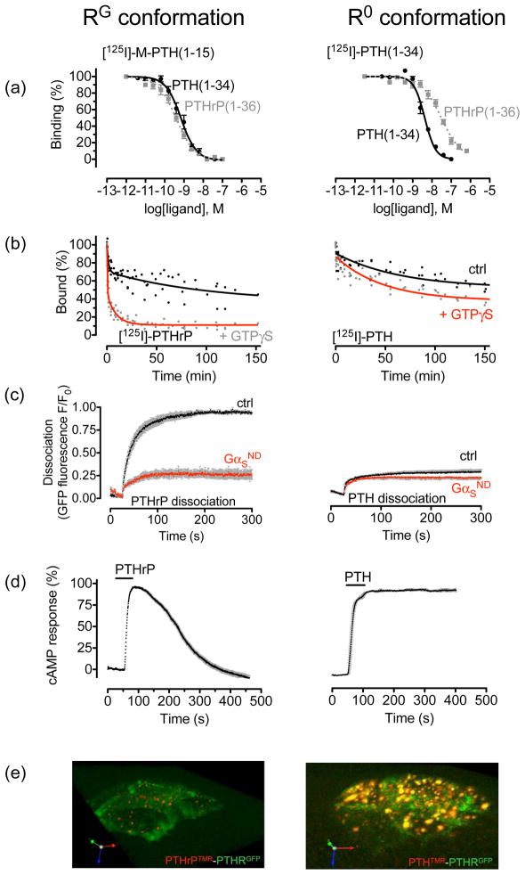
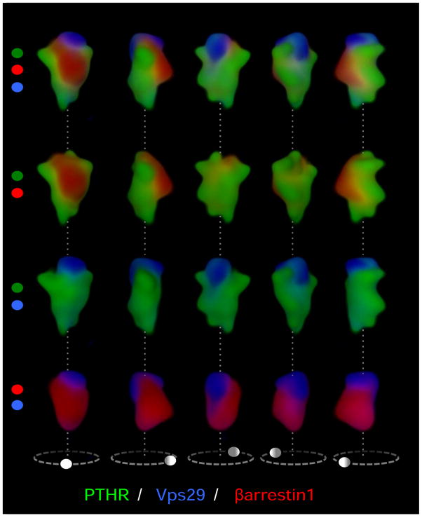
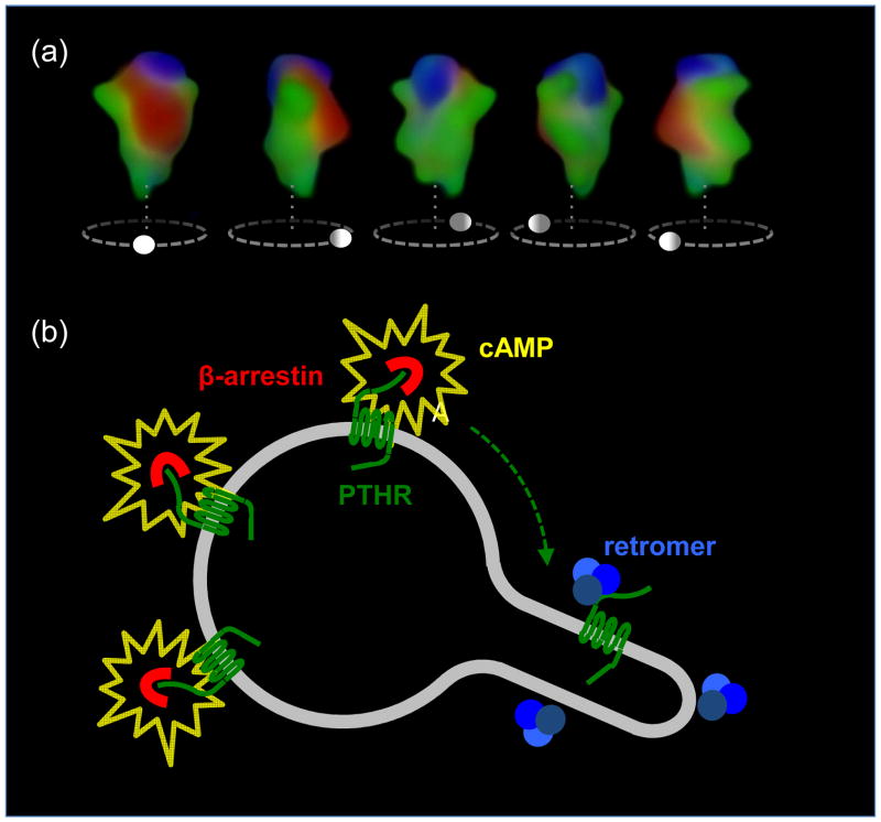
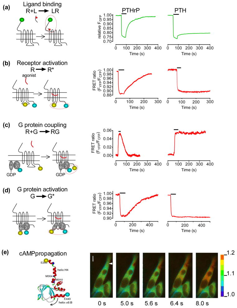
The classical model of arrestin-mediated desensitization of cell-surface G-protein-coupled receptors (GPCRs) is thought to be universal. However, this paradigm is incompatible with recent reports that the parathyroid hormone (PTH) receptor (PTHR), a crucial GPCR for bone and mineral ion metabolism, sustains G(S) activity and continues to generate cAMP for prolonged periods after ligand washout; during these periods the receptor is observed mainly in endosomes, associated with the bound ligand, G(S) and β-arrestins. In this review we discuss possible molecular mechanisms underlying sustained signaling by the PTHR, including modes of signal generation and attenuation within endosomes, as well as the biological relevance of such non-canonical signaling.


Conflict of interest statement

Conflicts of interest. J.-P.V. holds a patent on the FRET technology of measuring GPCR activation/deactivation by FRET (EP 1581811B1; US8084575).

Figures

Figure 1
Figure 1
PTHR conformations. (a–b) Cell membrane binding assays. Binding to the R0 and RG conformations of the PTHR are determined by competition reactions. For R0, [125 I]-PTH(—34) is used as a tracer radioligand and including GTPγS in the reaction; for RG, [125I]-PTH(—15) is used as a radioligand in the presence of a high-affinity, negative-dominant GαS subunit (Gαs-ND). (c–e) Life cells FRET-based assays. Averaged dissociation time courses of TMR-labeled ligands, PTH(–34)TMR (right panel) and PTHrP(–36)TMR (left panel), from GFP-tagged PTHR, GFPN-PTHR, are shown in the absence or presence of a Gαs-ND. FRET recordings from HEK-293 cells are shown as normalized ratios (c). Average time-courses of cAMP production in response to PTHrP(–36) (left) and PTH(1–84) (right) in HEK-293 cells stably expressing PTHR and co-transfected with the cAMP biosensor, EpacCFP/YFP. Individual cells were continuously perfused with buffer or with the hormone for the time indicated by the horizontal bar (d). A 3D view of tetramethylrhodamine (TMR)-labeled peptides, and a PTHR N-terminally tagged with GFP (GFPN-PTHR) in live HEK-293 cells by spinning disc confocal microscopy 30 min after ligand wash out. PTH(–34)TMR (red) and GFPN-PTHR (green) co-localized within endocytic compartments (right). In contrast, PTHrP(–36)TMR alone is detected as small puntae at internalized sites (left) (e). Adapted from (25, 33).
Figure 2
Figure 2
Signaling dynamics of PTHR on the early endosome. (a) We have recently shown that complexes of β-arrestin 1 (red) and PTHR (green) internalize to a compartment of the early endosome that is labeled red-green in a 3D reconstruction of early endosomes visualized with a spectral confocal microscope (top). A second compartment labeled with the sorting complex retromer (blue) is labeled blue-green. Arrestin and retromer do not colocalize, indicating that the two proteins localize in distinct compartments of the endosome, most likely the bulk domain (arrestin) and a domain dedicated to endosome-to-Golgi retrograde traffic (retromer). (b) Persistent complexes of PTHR-arrestin generate cAMP (yellow) from endosomal membranes. However, after arrestin-receptor decoupling, PTHR is free to bind retromer and sort to a compartment that does not support cAMP generation. Retromer-bound inactive PTHR then sorts to the trans-Golgi network before recycling to the plasma membrane. Adapted from (26).
Figure 3
Figure 3
Proposed model sustained PTHR signaling. Left panel, PTH-activated PTHR (green) generating cAMP (grey) by activation of adenyly cyclases internalizes to early endosomes in a process that involves binding of β-arrestin (red). Activated PTHR is then maintained in the early endosome bulk compartment by arrestin binding, where arrestin-mediated activation of ERK1/2 signaling causes inhibition of phosphodiesterases and permits sustained cAMP signaling. Right panel, Binding of PTHR and retromer (blue) causes sorting of the receptor to retrograde trafficking domains. Generation of cAMP is stopped after PTHR–retromer binding in the retrograde domain and retromer-mediated PTHR traffic to the Golgi. Adapted from (26).
Figure I
Figure I
Kinetics of PTHR signaling. (a) Ligand/receptor interaction measured by FRET between GFP-tagged PTHR and tetramethylrhodamine-labeled PTH(–34) or PTHrP(–36). Shown are the changes of GFP emission by FRET in response to rapid superfusion of diverse concentrations of ligand-TMR. (b) Following ligand application (horizontal bar), activation of PTHR was monitored in a single HEK-293 cell by a decrease in the FRET signal of PTHRCFP/YFP defined as the ratio of emission intensities of YFP/CFP. (c) The interaction between PTHR and GS proteins in response to ligand binding is measured as an increase in FRET between YFP-labeled PTHR and CFP-labeled Gγ2 in combination with GαS and Gβ1 proteins. (d) Detection of GS activation in cells expressing the wild-type PTHR by recording FRET between YFP-labeled Gαs and CFP-labeled Gγ2-subunits. (e) PTH-mediated cAMP response upon PTHR activation in HEK-293 cells measured as a decrease of FRET in the EpacCFP/YFP sensor. The panels show the propagation of the cAMP response represented as pseudocolored image of the FRET (CFP/YFP emission) ratio before and after stimulation of a single cell with PTH(–34) via a pipette indicated by an arrow at t = 0 s. The scale bar on the right indicates the pseudocolored scale of the fluorescence ratios. The inner bar represents 5 μm. Adapted from (25).

References

    1. Juppner H, Abou-Samra AB, Freeman M, Kong XF, Schipani E, Richards J, Kolakowski LF, Jr, Hock J, Potts JT, Jr, Kronenberg HM, et al. A G protein-linked receptor for parathyroid hormone and parathyroid hormone-related peptide. Science. 1991;254:1024–1026. - PubMed
    1. Schwindinger WF, Fredericks J, Watkins L, Robinson H, Bathon JM, Pines M, Suva LJ, Levine MA. Coupling of the PTH/PTHrP receptor to multiple G-proteins. Direct demonstration of receptor activation of Gs, Gq/11, and Gi(1) by [alpha-32P]GTP-gamma-azidoanilide photoaffinity labeling. Endocrine. 1998;8:201–209. - PubMed
    1. Gensure RC, Gardella TJ, Juppner H. Parathyroid hormone and parathyroid hormone-related peptide, and their receptors. Biochem Biophys Res Commun. 2005;328:666–678. - PubMed
    1. Singh AT, Gilchrist A, Voyno-Yasenetskaya T, Radeff-Huang JM, Stern PA. G alpha12/G alpha13 subunits of heterotrimeric G proteins mediate parathyroid hormone activation of phospholipase D in UMR-106 osteoblastic cells. Endocrinology. 2005;146:2171–2175. - PubMed
    1. Gesty-Palmer D, Chen M, Reiter E, Ahn S, Nelson CD, Wang S, Eckhardt AE, Cowan CL, Spurney RF, Luttrell LM, Lefkowitz RJ. Distinct beta-arrestin- and G protein-dependent pathways for parathyroid hormone receptor-stimulated ERK1/2 activation. J Biol Chem. 2006;281:10856–10864. - PubMed