. 2012 Aug 9;120(6):1254-61.
doi: 10.1182/blood-2012-02-410407. Epub 2012 Jun 18.

The folliculin-FNIP1 pathway deleted in human Birt-Hogg-Dubé syndrome is required for murine B-cell development

Affiliations

The folliculin-FNIP1 pathway deleted in human Birt-Hogg-Dubé syndrome is required for murine B-cell development

Masaya Baba et al. Blood. .

Abstract

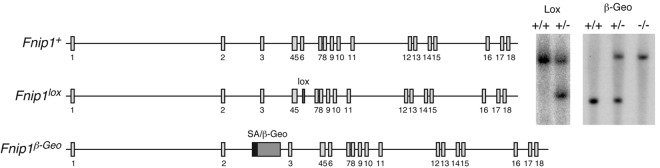
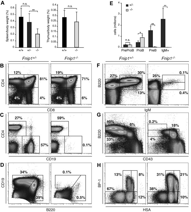
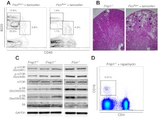
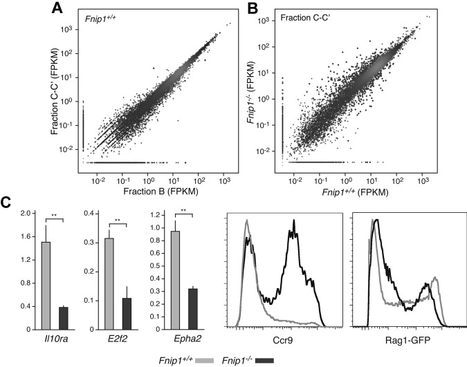
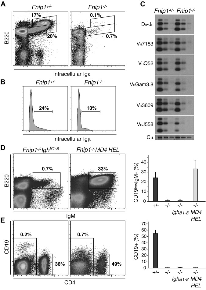
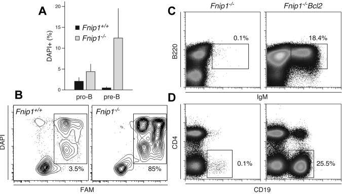
Birt-Hogg-Dubé (BHD) syndrome is an autosomal dominant disorder characterized by cutaneous fibrofolliculomas, pulmonary cysts, and kidney malignancies. Affected individuals carry germ line mutations in folliculin (FLCN), a tumor suppressor gene that becomes biallelically inactivated in kidney tumors by second-hit mutations. Similar to other factors implicated in kidney cancer, FLCN has been shown to modulate activation of mammalian target of rapamycin (mTOR). However, its precise in vivo function is largely unknown because germ line deletion of Flcn results in early embryonic lethality in animal models. Here, we describe mice deficient in the newly characterized folliculin-interacting protein 1 (Fnip1). In contrast to Flcn, Fnip1(-/-) mice develop normally, are not susceptible to kidney neoplasia, but display a striking pro-B cell block that is entirely independent of mTOR activity. We show that this developmental arrest results from rapid caspase-induced pre-B cell death, and that a Bcl2 transgene reconstitutes mature B-cell populations, respectively. We also demonstrate that conditional deletion of Flcn recapitulates the pro-B cell arrest of Fnip1(-/-) mice. Our studies thus demonstrate that the FLCN-FNIP complex deregulated in BHD syndrome is absolutely required for B-cell differentiation, and that it functions through both mTOR-dependent and independent pathways.


Figures

Figure 1
Figure 1
Generation of Fnip1 KO mice. Left schematics show Fnip1 WT (Fnip1+), targeted (Fnip1lox), and gene-trapped (Fnip1β-Geo) alleles. The Fnip1lox mutation was introduced in the germ line by targeting a neomycin resistance cassette (flanked by LoxP sites) within intron 5, followed by a second LoxP site within intron 6. The Neo-exon 6 sequence was removed by crossing targeted mice with β-actin-Cre transgenic mice. Exon 6 deletion generates a premature termination codon in exon 7. Fnip1β-Geo mice were derived from GeneTrap ES cell clone RRM154, which carries a splice acceptor (SA) β-Geo cassette inserted within intron 2. Proper gene targeting in both cases was confirmed by Southern blotting (right gel pictures).
Figure 2
Figure 2
Severe pro-B cell arrest in Fnip1−/− mice. (A) Left bar graph: spleen weight (given as percentage of body weight) in Fnip1−/− mice relative to littermate controls graphed as the mean ± SD; NS = no statistical significance, **P < .01 (Student t test), n = 9 (Fnip1+/+), 17 (Fnip1+/−), and 13 (Fnip1−/−). Right graph shows the same for thymuses. n = 6 (Fnip1+/+), and 8 (Fnip1−/−). (B-D) Fluorocytometric analysis of CD4+ and CD8+ thymocytes (B), CD4+ and CD19+ splenocytes (C), and B1 (B220highCD19low) and B2 (B220lowCD19high) peritoneal cavity B cells (D). (E) Absolute number of pre–pro-B (B220+IgMCD19), pro-B (B220+IgMCD19+CD43+CD25), pre-B (B220+IgMCD19+CD43CD25+), and immature (B220+IgM+) B cells in BM of Fnip1+/− and Fnip1−/−mice. *P < .05, **P < .01 (Student t test), n = 4 for both strains. (F-H) Fluorocytometric analysis of B220 and IgM (F), B220 and CD43 (G), and BP-1 and HSA (H) expression in BM cells from Fnip1+/−and Fnip1−/−mice. Each analysis is representative of at least 3 independent experiments.
Figure 3
Figure 3
Comparison of Fnip1−/− and tamoxifen-induced Flcn deficient mice. (A) BM B-cell analysis in Flcnf/+ERCre+ and Flcnf/−ERCre+ mice 4 weeks posttamoxifen (TM) injection. Cell samples were stained with CD43 (FITC) and B220 (PE). (B) Flcnf/−ERCre+ mice treated with tamoxifen (right micrograph) show hyperplastic cystic lesions (denoted with arrowheads) in kidney. Conversely, Fnip−/− mice rarely display cysts (left micrograph). Scale bar = 200 μm. (C) Western blot analysis of kidney cell lysates from Fnip1+/−, Fnip1−/−, and Flcn−/− mice. mTOR activation was assessed via phosphorylation of mTOR serines 2481 and 2448, as well as phosphorylation of ribosomal protein S6, serines 240/244 and 235/236. GAPDH expression was used as a loading control. (D) Failure to rescue peripheral CD19+ B lymphocytes in Fnip1−/− mice treated with buffer or rapamycin (2 mg/kg) for 3 weeks. Each fluorescence-activated cell sorter (FACS) analysis is representative of at least 3 independent experiments.
Figure 4
Figure 4
Transcriptome analysis of Fnip1−/− pro-B cells. (A) Scatter plot showing a comparative analysis between Fnip1+/+ transcriptomes (mRNA-Seq FPKM values) obtained from the Hardy pro-B cell fractions B and C-C′. Red dots represent genes whose transcription is statistically different between the 2 fractions; n = 1 (using pooled estimates of dispersion). (B) Same analysis as in panel A but comparing fractions C-C′ between Fnip1+/+ and Fnip1−/− mice; n = 2 for each genotype. (C) Bar graphs showing RT-qPCR measurements of transcription in Fnip1+/+ and Fnip1−/− pro-B cells from selected genes (n = 3). Histograms showing ccr9 and Rag1-GFP expression, as determined by flow cytometry, on B220+IgMCD25 pro-B cells from KO and control mice. Each FACS analysis is representative of at least 3 independent experiments.
Figure 5
Figure 5
Prerecombined antibody genes do not rescue Fnip1−/− B cells. (A) Igκ staining in Fnip1+/− and Fnip1−/− BM B220+ B cells. (B) Histogram of intracellular Igμ expression in B220+CD43+ pro-B cells from Fnip1+/− and Fnip1−/− mice. (C) D-J and V-DJ recombination analysis in WT and KO fraction C/C′ B cells as determined by PCR-Southern blot. (D) B220/IgM expression profiles in Fnip1−/− mice expressing B1-8 heavy chain (IghB18) or the αHEL heavy and light chain transgene (MD4 HEL). Right bar graph represents the mean ± SD of IgM+B220+ B cells from each strain; n values were as follows: 6 for Fnip1+/− and Fnip1−/−, and 3 for Fnip1−/−IghB1-8 and Fnip1−/−MD4 HEL. (E) Same analysis as in panel D but using CD19 and CD4 staining of splenocytes; n values: 5 mice for Fnip1+/− and Fnip1−/−, and 4 mice for Fnip1−/−IghB1-8 and Fnip1−/−MD4 HEL. Each FACS analysis is representative of at least 3 independent experiments.
Figure 6
Figure 6
Increased caspase activation and apoptosis in Fnip1−/− B cells. (A) Bar graph showing the frequency of dead cells within pro-B (B220lowIgMCD25) and pre-B (B220lowIgMCD25+) populations from Fnip1+/+ and Fnip1−/− mice as measured by DAPI staining. Mean ± SD from 4 independent experiments, (pro-B) P = .1, (pre-B) P < .02, Student t test. (B) Flow cytometric measurement of caspase activity in Fnip1+/+ and Fnip1−/− pre-B cells from interleukin 7 ex vivo cultures. Twenty-four hours after interleukin 7 removal cells were stained with DAPI and FAM, a carboxyfluorescein moiety that becomes covalently linked to the active site of caspases. (C) Flow cytometric analysis of BM and (D) spleen cells from Fnip1−/− mice reconstituted with an Eμ-Bcl2 transgene. BM cells were stained with anti-B220 and anti-IgM antibodies; staining for splenocytes was with anti-CD4 and anti-CD19. Each FACS analysis is representative of 3 independent experiments.

References

    1. Schmidt LS, Nickerson ML, Warren MB, et al. Germline BHD-mutation spectrum and phenotype analysis of a large cohort of families with Birt-Hogg-Dube syndrome. Am J Hum Genet. 2005;76(6):1023–1033. - PMC - PubMed
    1. Nickerson ML, Warren MB, Toro JR, et al. Mutations in a novel gene lead to kidney tumors, lung wall defects, and benign tumors of the hair follicle in patients with the Birt-Hogg-Dube syndrome. Cancer Cell. 2002;2(2):157–164. - PubMed
    1. Okimoto K, Kouchi M, Matsumoto I, Sakurai J, Kobayashi T, Hino O. Natural history of the Nihon rat model of BHD. Curr Mol Med. 2004;4(8):887–893. - PubMed
    1. Vocke CD, Yang Y, Pavlovich CP, et al. High frequency of somatic frameshift BHD gene mutations in Birt-Hogg-Dube-associated renal tumors. J Natl Cancer Inst. 2005;97(12):931–935. - PubMed
    1. Hasumi Y, Baba M, Ajima R, et al. Homozygous loss of BHD causes early embryonic lethality and kidney tumor development with activation of mTORC1 and mTORC2. Proc Natl Acad Sci U S A. 2009;106(44):18722–18727. - PMC - PubMed

Publication types

MeSH terms