. 2013;22(1):133-45.
doi: 10.3727/096368912X647162. Epub 2012 Jun 15.

Insulin-producing cells from adult human bone marrow mesenchymal stem cells control streptozotocin-induced diabetes in nude mice

Affiliations

Insulin-producing cells from adult human bone marrow mesenchymal stem cells control streptozotocin-induced diabetes in nude mice

Mahmoud M Gabr et al. Cell Transplant. 2013.

Abstract

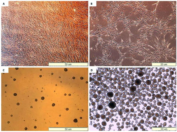
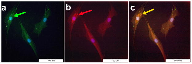
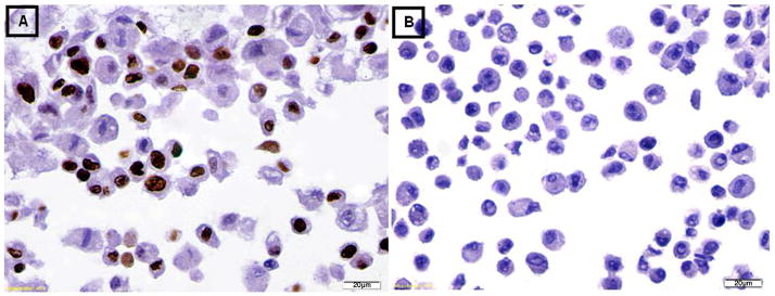
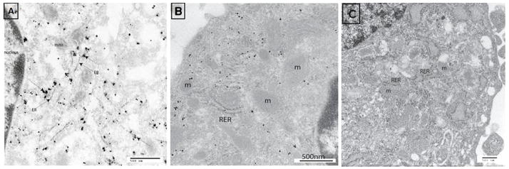
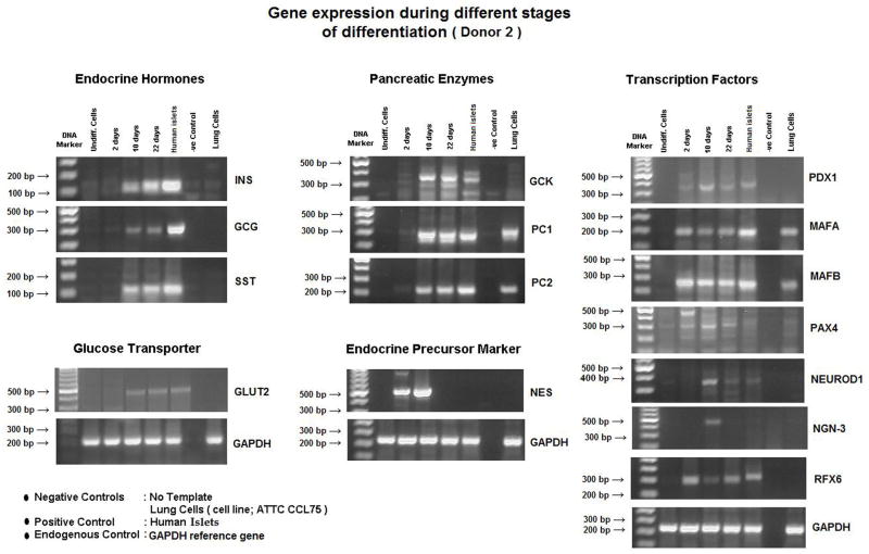
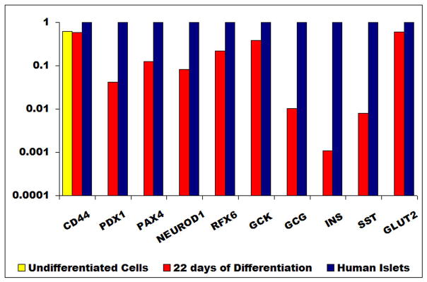
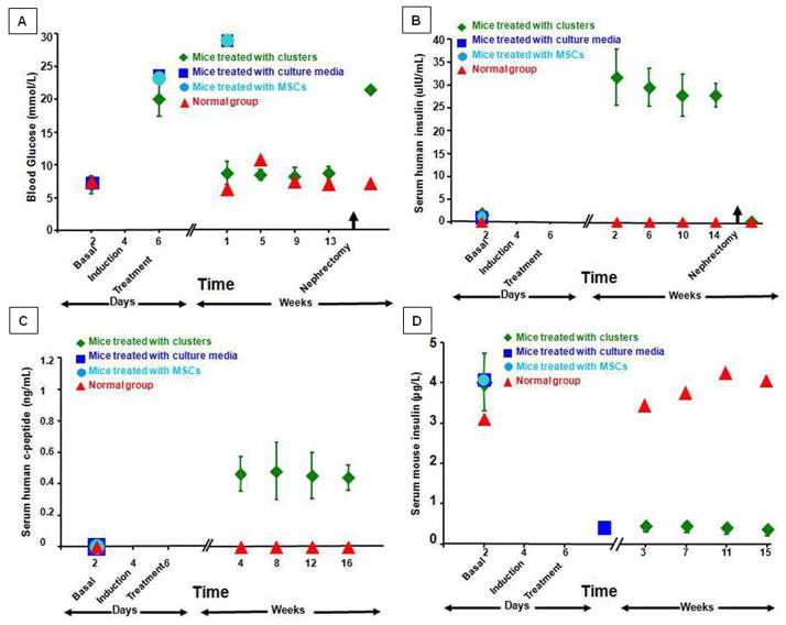
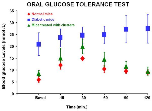
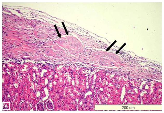
Harvesting, expansion, and directed differentiation of human bone marrow-derived mesenchymal stem cells (BM-MSCs) could provide an autologous source of surrogate β-cells that would alleviate the limitations of availability and/or allogenic rejection following pancreatic or islet transplantation. Bone marrow cells were obtained from three adult type 2 diabetic volunteers and three nondiabetic donors. After 3 days in culture, adherent MSCs were expanded for two passages. At passage 3, differentiation was carried out in a three-staged procedure. Cells were cultured in a glucose-rich medium containing several activation and growth factors. Cells were evaluated in vitro by flow cytometry, immunolabeling, RT-PCR, and human insulin and c-peptide release in responses to increasing glucose concentrations. One thousand cell clusters were inserted under the renal capsule of diabetic nude mice followed by monitoring of their diabetic status. At the end of differentiation, ∼5-10% of cells were immunofluorescent for insulin, c-peptide or glucagon; insulin, and c-peptide were coexpressed. Nanogold immunolabeling for electron microscopy demonstrated the presence of c-peptide in the rough endoplasmic reticulum. Insulin-producing cells (IPCs) expressed transcription factors and genes of pancreatic hormones similar to those expressed by pancreatic islets. There was a stepwise increase in human insulin and c-peptide release by IPCs in response to increasing glucose concentrations. Transplantation of IPCs into nude diabetic mice resulted in control of their diabetic status for 3 months. The sera of IPC-transplanted mice contained human insulin and c-peptide but negligible levels of mouse insulin. When the IPC-bearing kidneys were removed, rapid return of diabetic state was noted. BM-MSCs from diabetic and nondiabetic human subjects could be differentiated without genetic manipulation to form IPCs that, when transplanted, could maintain euglycemia in diabetic mice for 3 months. Optimization of the culture conditions are required to improve the yield of IPCs and their functional performance.


Conflict of interest statement

Conflicts of interest:

The authors disclose no Conflicts of interest

Figures

Figure 1
Figure 1. Morphological changes of MSCs during differentiation
(A) Undifferentiated MSCs, at the end of the expansion phase. (B) Formation of cell aggregates at day 10. (C) Forrmation of cell clusters at day 18. (D) Collected clusters.
Figure 2
Figure 2. Immunoflourescent staining of differentiated IPCS
(A) Positive staining for insulin (green) with counter staining for DAPI (arrow). (B) Positive staining for c-peptide (red) with counter staining for DAPI (arrow). (C) Electronic merge of insulin and c-peptide. Co-expression of insulin and c-peptide (yellow) by the same cells could be seen (arrow).
Figure 3
Figure 3. Immunohistochemical staining for the proliferation marker Ki-67
(A) The undifferentiated MSCs were positive for staining with Ki-67. (B) The differentiated IPCs were negative for staining with Ki-67.
Figure 4
Figure 4. Immuno-staining with nanogold
(A) Ultrastructure analysis of the differentiated IPCs shows concentration of c-peptide (arrows) at the cisternae of rough endoplasmic reticulum. (B) Fragment of undifferentiated cell shows unlabeled cytoplasm for c-peptide (negative control). (C) Fragment of pancreatic islet cells shows c-peptide labeling in the vicinity of rough endoplasmic reticulum (positive control). RER: Rough endoplasmic reticulum. m: Mitochondria.
Figure 5
Figure 5. Gene expression profile
(A) RT-PCR of the undifferentiated MSCs and during the various phases of their differentiation (days 2,10,22). At the end of differentiation (day 22), the gene expression of IPCs was similar to that of human islets. Both expressed transcription factors (PDX-1, MafA, MafB, Pax4, Rfx6 and neuro D-1), pancreatic enzymes (GK, PC1 & PC2) and endocrine hormones (insulin, glucagon and somatostation). (B) qPCR : undifferentiated cells did not express any transcription factors or genes of endocrine hormones. At full differentiation (22 days), PDX-1 expression was between 1 and 10% of that of human islets. Insulin gene expression was < 1% of that of human islets.
Figure 6
Figure 6. Biochemical profile of diabetic nude mice treated by implantation of IPCs under the renal capsule (green)
The blood glucose levels (a) were normalized few days after implantation and maintained throughout the observation period. The diabetic status resumed following nephrectomy of the IPCs-bearing kidneys. Serum human insulin (b) and c-peptide (c) became measurable after implantation and rapidly disappeared following nephrectomy of the IPCs-bearing kidneys. On the other hand, serum mouse insulin (d) was negligible.
Figure 7
Figure 7
The oral glucose tolerance tests. transplanted animals (green), normal animals (red) and diabetic animals (blue). The curve of the IPCs-transplanted animals approximated that of normal controls.
Figure 8
Figure 8. Histology of the removed IPCs-bearing kidneys
Hematoxylin and Eosin examination (A) demonstrates the implanted IPCs under the renal capsule (arrows). Positive. Immunoflourescent staining for insulin (arrow) (A) and c-peptide (arrow) (C). Electronic merging reveals co-expression of insulin and c-peptide by the same cells (arrow) (D). Confocal study with dual labeling for insulin (red) and glucagon (green) was positive in the IPCs under the renal capsule (E) and in human islets (F). These two hormones were localized in different cell populations (arrows).

Similar articles

Cited by

References

    1. Bilinski SM, Jaglarz MK, Dougherty MT, Kloc M. Electron microscopy, immunostaining, cytoskeleton visualization, in situ hybridization, and three-dimensional reconstruction of Xenopus occytes. Methods. 2010;51(1):11–19. - PubMed
    1. Bonner-Weir S, Deery D, Leahy JL, Weir GC. Compensatory growth of pancreatic beta-cells in adult rats after short-term glucose infusion. Diabetes. 1989;38(1):49–53. - PubMed
    1. Calne R, Ghoneim MA. Novel diabetes therapy: The seven pillars of credibility. Treatment Strategies Diabetes. Available online at: www.treatmentstrategies.co.uk./Diabetes.html.
    1. Calne RY, Gan SU, Lee KO. Stem cell and gene therapies for diabetes mellitus. Nat Rev Endocrinol. 2010;6(3):173–177. - PubMed
    1. Chatterjee AK, Sieradzki J, Schatz H. Epidermal growth factor stimulates (pro-) insulin biosynthesis and 3H-thymidine incorporation in isolated pancreatic rat islets. Horm Metab Res. 1986;18(12):873–874. - PubMed

Publication types