

Orexinergic signaling mediates light-induced neuronal activation in the dorsal raphe nucleus
- PMID: 22710065
- PMCID: PMC3412924
- DOI: 10.1016/j.neuroscience.2012.06.020
Orexinergic signaling mediates light-induced neuronal activation in the dorsal raphe nucleus
Abstract
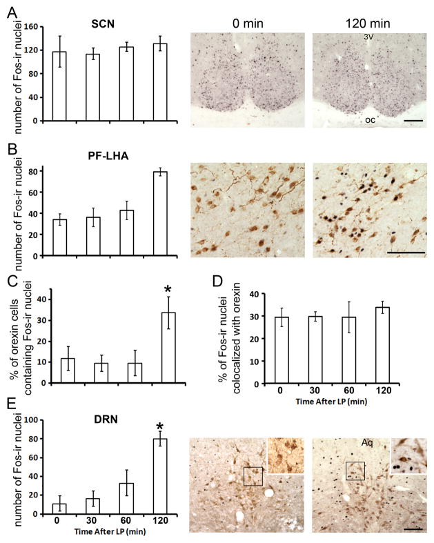
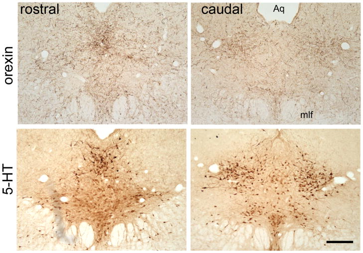
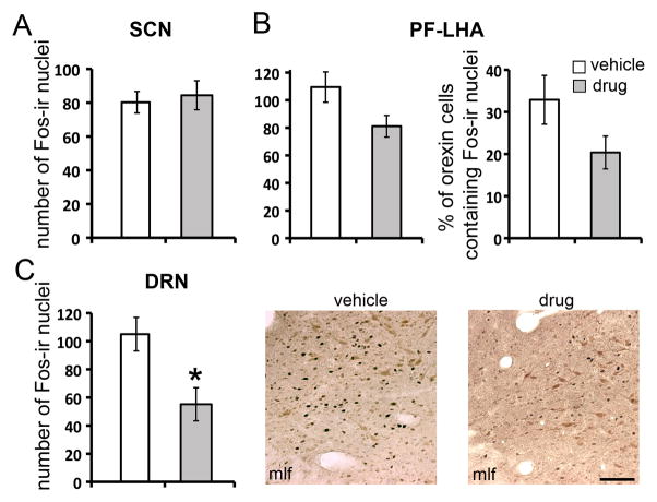
Seasonal affective disorder (SAD), a major depressive disorder recurring in the fall and winter, is caused by the reduction of light in the environment, and its depressive symptoms can be alleviated by bright light therapy. Both circadian and monoaminergic systems have been implicated in the etiology of SAD. However, the underlying neural pathways through which light regulates mood are not well understood. The present study utilized a diurnal rodent model, Arvicanthis niloticus, to explore the neural pathways mediating the effects of light on brain regions involved in mood regulation. Animals kept in constant darkness received light exposure in early subjective day, the time when light therapy is usually applied. The time course of neural activity following light exposure was assessed using Fos protein as a marker in the following brain regions/cells: the suprachiasmatic nucleus (SCN), orexin neurons in the perifornical-lateral hypothalamic area (PF-LHA) and the dorsal raphe nucleus (DRN). A light-induced increase in Fos expression was observed in orexin neurons and the DRN, but not in the SCN. As the DRN is densely innervated by orexinergic inputs, the involvement of orexinergic signaling in mediating the effects of light on the DRN was tested in the second experiment. The animals were injected with the selective orexin receptor type 1 (OXR1) antagonist SB-334867 prior to the light exposure. The treatment of SB-334867 significantly inhibited the Fos induction in the DRN. The results collectively point to the role of orexin neurons in mediating the effects of light on the mood-regulating monoaminergic areas, suggesting an orexinergic pathway that underlies light-dependent mood fluctuation and the beneficial effects of light therapy.
Copyright © 2012 IBRO. Published by Elsevier Ltd. All rights reserved.
Figures
References
-
- Abrahamson EE, Leak RK, Moore RY. The suprachiasmatic nucleus projects to posterior hypothalamic arousal systems. Neuroreport. 2001;12:435–440. - PubMed
-
- Ashkenazy-Frolinger T, Kronfeld-Schor N, Juetten J, Einat H. It is darkness and not light: Depression-like behaviors of diurnal unstriped Nile grass rats maintained under a short photoperiod schedule. J Neurosci Methods. 2010;186:165–170. - PubMed
-
- Backberg M, Hervieu G, Wilson S, Meister B. Orexin receptor-1 (OX-R1) immunoreactivity in chemically identified neurons of the hypothalamus: focus on orexin targets involved in control of food and water intake. Eur J Neurosci. 2002;15:315–328. - PubMed
-
- Blanchong JA, McElhinny TL, Mahoney MM, Smale L. Nocturnal and diurnal rhythms in the unstriped Nile rat, Arvicanthis niloticus. J Biol Rhythms. 1999;14:364–377. - PubMed
-
- Burt J, Alberto CO, Parsons MP, Hirasawa M. Local network regulation of orexin neurons in the lateral hypothalamus. Am J Physiol Regul Integr Comp Physiol. 2011;301:R572–580. - PubMed
Publication types
MeSH terms
Substances
Grants and funding
LinkOut - more resources
Full Text Sources
Research Materials
