Review
. 2012 Aug;33(3):230-51.
doi: 10.1016/j.yfrne.2012.06.002. Epub 2012 Jun 16.

Neurodevelopmental effects of insulin-like growth factor signaling

Affiliations
Review

Neurodevelopmental effects of insulin-like growth factor signaling

John O'Kusky et al. Front Neuroendocrinol. 2012 Aug.

Abstract

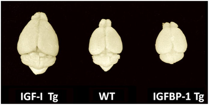
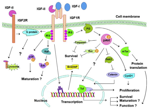
Insulin-like growth factor (IGF) signaling greatly impacts the development and growth of the central nervous system (CNS). IGF-I and IGF-II, two ligands of the IGF system, exert a wide variety of actions both during development and in adulthood, promoting the survival and proliferation of neural cells. The IGFs also influence the growth and maturation of neural cells, augmenting dendritic growth and spine formation, axon outgrowth, synaptogenesis, and myelination. Specific IGF actions, however, likely depend on cell type, developmental stage, and local microenvironmental milieu within the brain. Emerging research also indicates that alterations in IGF signaling likely contribute to the pathogenesis of some neurological disorders. This review summarizes experimental studies and shed light on the critical roles of IGF signaling, as well as its mechanisms, during CNS development.


Figures

Fig. 1
Fig. 1
Brains from an IGF-IMT-I overexpressing Tg mouse (IGF-I Tg), a wild type control mouse (WT), and an IGFBP-1 Tg mouse that ectopically express brain IGFBP-1, which reduces IGF availability.
Fig. 2
Fig. 2
Representative sections through the head and brain of an E16 homozygous igf1rNestin-KO embryo (right panels) and its heterozygous littermate (left panels), stained with hematoxylin and eosin. In heterozygous embryos the brain exhibits relatively normal morphology at all levels, although smaller in size compared to littermate controls (not illustrated). In homozygous embryos the brain exhibits gross malformations in some mice. There is a failure to close the longitudinal suture of the skull in homozygous embryos, accompanied by the extrusion of gray matter through the skull and sloping forward (Panels B and C, arrows). In heterozygous embryos the developing hippocampus and DG are located along the medial edge of the telencephalic wall as in controls (E, arrow). In homozygous embryos the hippocampus and DG rotated to the lateral edge of the telencephalic wall (E, arrows) following extensive apoptosis in the dorsolateral wall at E14 and protrusion of the underlying telencephalic structures.
Fig. 3
Fig. 3
Representative sections of the right cerebral hemisphere in a control embryo (A), a heterozygous igf1rNestin-KO embryo (B), and a homozygous igf1rNestin-KO embryo at E16.
Fig. 4
Fig. 4
Immunostaining for GAD67, calbindin, and calretinin in the ventral prefrontal cortex of an adult heterozygous igf1rNestin-KO mouse (B, D, and F) and its littermate control (A, C, and E). Coronal sections through the ventral prefrontal cortex have been stained with antibodies against GAD67 (A and B), calbindin (C and D) and calretinin (E and F). Note the decreased density of GAD67-immunoreactive GABAergic neurons in (B) and the decreased density of calbindin-immunoreactive GABAergic neurons in (D) with no change in the density of calretinin-immunoreactive GABAergic neurons in (F), when compared to controls.
Fig. 5
Fig. 5
IGF signaling in the CNS. In this largely simplified diagram the IGF signaling pathways in the CNS are schematically depicted. Other signaling molecules and pathways described in non-neural cells systems are not included in the Figure, although it is possible that they are also involved. ⊥ = inhibitory modification, and ↓ = stimulatory modification.

Similar articles

Cited by

References

    1. Aberg MA, Aberg ND, Hedbacker H, Oscarsson J, Eriksson PS. Peripheral infusion of IGF-I selectively induces neurogenesis in the adult rat hippocampus. J Neurosci. 2000;20:2896–2903. - PMC - PubMed
    1. Aberg MA, Aberg ND, Palmer TD, Alborn AM, Carlsson-Skwirut C, Bang P, Rosengren LE, Olsson T, Gage FH, Eriksson PS. IGF-I has a direct proliferative effect in adult hippocampal progenitor cells. Mol Cell Neurosci. 2003a;24:23–40. - PubMed
    1. Aberg ND, Blomstrand F, Aberg MA, Bjorklund U, Carlsson B, Carlsson-Skwirut C, Bang P, Ronnback L, Eriksson PS. Insulin-like growth factor-I increases astrocyte intercellular gap junctional communication and connexin43 expression in vitro. J Neurosci Res. 2003b;74:12–22. - PubMed
    1. Aberg ND, Johansson UE, Aberg MA, Hellstrom NA, Lind J, Bull C, Isgaard J, Anderson MF, Oscarsson J, Eriksson PS. Peripheral infusion of insulin-like growth factor-I increases the number of newborn oligodendrocytes in the cerebral cortex of adult hypophysectomized rats. Endocrinology. 2007;148:3765–3772. - PubMed
    1. Abuzzahab MJ, Schneider A, Goddard A, Grigorescu F, Lautier C, Keller E, Kiess W, Klammt J, Kratzsch J, Osgood D, Pfaffle R, Raile K, Seidel B, Smith RJ, Chernausek SD. IGF-I receptor mutations resulting in intrauterine and postnatal growth retardation. New Engl J Med. 2003;349:2211–2222. - PubMed

Publication types

MeSH terms

Substances