Review
. 2012 Jun 19;8(7):380-90.
doi: 10.1038/nrneurol.2012.99.

Neuronal autoantigens--pathogenesis, associated disorders and antibody testing

Affiliations
Review

Neuronal autoantigens--pathogenesis, associated disorders and antibody testing

Eric Lancaster et al. Nat Rev Neurol. .

Abstract

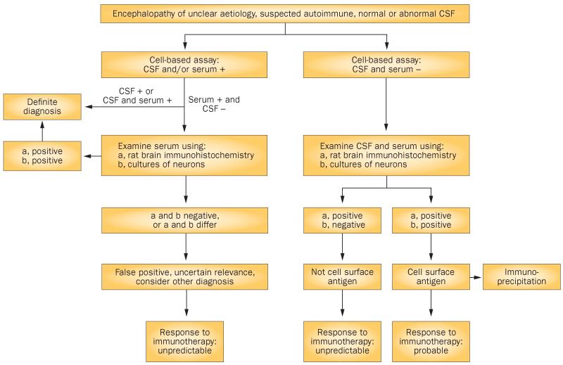
The discovery of disorders that are associated with antibodies to neuronal cell-surface proteins has led to a paradigm shift in our understanding of CNS autoimmunity. These disorders can occur in patients with or without cancer-often children or young adults who develop psychosis, catatonic or autistic features, memory problems, abnormal movements, or seizures that were previously considered idiopathic. The autoantigens in such cases have crucial roles in synaptic transmission, plasticity and peripheral nerve excitability. Patients can be comatose or encephalopathic for months and yet fully recover with supportive care and immunotherapy. By contrast, disorders in which the antibodies target intracellular antigens, and in which T-cell-mediated irreversible neuronal degeneration occurs, show a considerably poorer response to treatment. In this article, we review the various targets of neuronal antibodies, focusing predominantly on autoantigens located on the cell surface or synapses-namely, N-methyl-D-aspartate receptors, α-amino-3-hydroxy-5-methyl-4-isoxazolepropionic acid receptors, γ-aminobutyric acid receptors, leucine-rich glioma-inactivated protein 1, contactin-associated protein-like 2, and metabotropic glutamate receptors. We also provide an algorithm to identify and assess antibodies that bind to cell-surface and synaptic antigens.


Figures

Figure 1
Figure 1
Autoantigens and mechanisms of neuronal dysfunction. a | Antibodies to intracellular antigens, such as HuD, might not be pathogenic but could instead indicate a T-cell-mediated response against neurons. b | Intracellular synaptic antigens, such as GAD65, may be targeted by both antibodies and T-cell-mediated mechanisms. Which of these mechanisms is more important remains controversial. c | Cell-surface receptors are functionally disrupted by antibodies. Antibodies against NMDAR and AMPAR have been shown to cause receptor cross-linking and internalization. Abbreviations: AMPAR, α-amino-3-hydroxy-5-methyl-4-isoxazolepropionic acid receptor; GAD65, 65 kDa glutamic acid decarboxylase; NMDAR, N-methyl-d-aspartate receptor.
Figure 2
Figure 2
Caspr2 interaction with juxtaparanodal proteins. Caspr2 is an axonal protein that binds contactin-2 in cis and trans orientations to organize the juxtaparanodal region. Caspr2 links to PDZ-binding proteins, and to the cytoskeleton via protein 4.1B. Caspr2 organizes Kv1 potassium channels in the juxtaparanodal region, although the underlying mechanism remains to be determined. Abbreviation: Caspr2, contactin-associated protein-like 2.
Figure 3
Figure 3
Algorithm for identification and assessment of antibodies to neuronal cell-surface antigens. Comprehensive assessment of known and novel antibodies to cell-surface antigens depends on examination of the reactivity of serum and CSF. Some patients may have normal results on routine CSF studies (for example, cell counts, protein levels and oligoclonal bands) but test positive for specific antibodies to neuronal cell-surface antigens (for example, leucine-rich glioma-inactivated protein 1 or the N-methyl-d-aspartate receptor) in the CSF. In patients with symptomatic encephalitis, we always test for antibodies in CSF, as antibodies can be present in or absent from serum. Cases with only low levels of antibodies in the CSF have been reported, although the relevance and identity of these antibodies are unclear. In such cases, confirmation that antibodies react with the cell surface of neurons (as tested in cell culture) and with the brain neuropil (as tested in rat brain slice preparations) is strongly recommended. Detection of a novel cell-surface reactivity should be followed by characterization of the antigen by immunoprecipitation and mass spectrometry. Abbreviation: CSF, cerebrospinal fluid.

References

    1. Dalmau J, et al. Paraneoplastic anti-N-methyl-d-aspartate receptor encephalitis associated with ovarian teratoma. Ann. Neurol. 2007;61:25–36. - PMC - PubMed
    1. Lai M, et al. AMPA receptor antibodies in limbic encephalitis alter synaptic receptor location. Ann. Neurol. 2009;65:424–434. - PMC - PubMed
    1. Lancaster E, et al. Antibodies to the GABAB receptor in limbic encephalitis with seizures: case series and characterisation of the antigen. Lancet Neurol. 2010;9:67–76. - PMC - PubMed
    1. Lai M, et al. Investigation of LGI1 as the antigen in limbic encephalitis previously attributed to potassium channels: a case series. Lancet Neurol. 2010;9:776–785. - PMC - PubMed
    1. Lancaster E, et al. Investigations of Caspr2, an autoantigen of encephalitis and neuromyotonia. Ann. Neurol. 2011;69:303–311. - PMC - PubMed

Publication types

MeSH terms