. 2013 Apr 18;32(16):2005-15.
doi: 10.1038/onc.2012.230. Epub 2012 Jun 18.

TGF-β stimulates Pyk2 expression as part of an epithelial-mesenchymal transition program required for metastatic outgrowth of breast cancer

Affiliations

TGF-β stimulates Pyk2 expression as part of an epithelial-mesenchymal transition program required for metastatic outgrowth of breast cancer

M K Wendt et al. Oncogene. .

Abstract

Epithelial-mesenchymal transition (EMT) programs are essential in promoting breast cancer invasion, systemic dissemination and in arousing proliferative programs in breast cancer micrometastases, a reaction that is partially dependent on focal adhesion kinase (FAK). Many functions of FAK are shared by its homolog, protein tyrosine kinase 2 (Pyk2), raising the question as to whether Pyk2 also participates in driving the metastatic outgrowth of disseminated breast cancer cells. In addressing this question, we observed Pyk2 expression to be (i) significantly upregulated in recurrent human breast cancers; (ii) differentially expressed across clonal isolates of human MDA-MB-231 breast cancer cells in a manner predictive for metastatic outgrowth, but not for invasiveness; and (iii) dramatically elevated in ex vivo cultures of breast cancer cells isolated from metastatic lesions as compared with cells that produced the primary tumor. We further show that metastatic human and murine breast cancer cells robustly upregulate their expression of Pyk2 during EMT programs stimulated by transforming growth factor-β (TGF-β). Genetic and pharmacological inhibition of Pyk2 demonstrated that the activity of this protein tyrosine kinase was dispensable for the ability of breast cancer cells to undergo invasion in response to TGF-β, and to form orthotopic mammary tumors in mice. In stark contrast, Pyk2-deficiency prevented TGF-β from stimulating the growth of breast cancer cells in 3D-organotypic cultures that recapitulated pulmonary microenvironments, as well as inhibited the metastatic outgrowth of disseminated breast cancer cells in the lungs of mice. Mechanistically, Pyk2 expression was inversely related to that of E-cadherin, such that elevated Pyk2 levels stabilized β1 integrin expression necessary to initiate the metastatic outgrowth of breast cancer cells. Thus, we have delineated novel functions for Pyk2 in mediating distinct elements of the EMT program and metastatic cascade regulated by TGF-β, particularly the initiation of secondary tumor outgrowth by disseminated cells.


Conflict of interest statement

Conflicts of interest

The authors declare no conflict of interest.

Figures
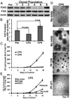
Figure 1
Figure 1
Elevated Pyk2 expression is associated with enhanced 3D-organotypic growth, but not with the invasiveness of MDA-231 cells. (a) Immunoblot analysis of parental (Par) and isolated clonal populations of MDA-MB-231 cells showed that Pyk2 expression was highly variable across these cell lines, while that of FAK was ubiquitous and equivalent. (b) Pyk2 expression inversely correlated with the invasive potential of MDA-231 clonal populations (CP) CP2 and CP4. Data are the mean (±SE) of 2 independent experiments completed in triplicate. (c) Robust Pyk2 expression in CP2 cells correlates with their enhanced 3D–outgrowth, an event that was lacking in Pyk2-deficient CP4 cells. (d) Photomicrographs of CP2 and CP4 cells propagated in 3D-organotypic cultures. Where indicated, CP2 cells were grown in the absence (No Inhib) or presence of the FAK specific inhibitor, PF573228 (1 µM; PF228), or the dual FAK/Pyk2 inhibitor, PF562271 (1 µM; PF271). (e) Bioluminescent quantification of CP2 proliferation in 3D–cultures shown in Panel d. Data for Panels c and e are the mean (±SE) of 3 independent experiments completed in triplicate.
Figure 2
Figure 2
TGF-β induces Pyk2 expression via Src and Smad4 signaling. (a) Human MDA-MB-231 breast cancer cells were propagated in traditional 2D–cultures (2D) or 3D-organotypic cultures (3D) for 5 days in the absence or presence of TGF-β1 (5 ng/ml) as indicated. Afterward, differences in the expression levels of Pyk2 and FAK were monitored by immunoblotting. The N-terminally-directed FAK antibody also readily detected the accumulation of the FAK FERM domain as shown. β-actin served as a loading control. (b and c) 4T07 and 4T1 cells were stimulated with TGF-β1 (5 ng/ml) over a span of 48 h as indicated. Afterward, changes in the levels of Pyk2 and FAK mRNA were quantified by real-time PCR (b), or by immunoblotting (c). Changes in β3 integrin (β3-Int) expression served as a surrogate marker for EMT, while β-actin served as a loading control. (d) 4T1 cells were treated for 0–72 h with the inhibitors against TβR-I (TBR1 or SBTBR) or Src (PP2). Afterward, changes in basal levels of Pyk2 expression were assessed by immunoblot analysis. (e) Dual-bioluminescent SBE reporter 4T1 cells were propagated in traditional 2D- or 3D-organotypic cultures for 4 days in the absence or presence of TGF-β1 (5 ng/ml) prior to monitoring firefly and renilla luciferase activity. Data are the mean (±SE) of 3 independent experiments completed in triplicate. (f) Control (scram) or Smad4-depleted (shSmad4) MDA-231-CP4 cells were stimulated with TGF-β1 (5 ng/ml) for 48 h. Smad4-depleted cells failed to upregulate Pyk2. Data in Panels a, c, d, and f are representative of at least 2 independent experiments, while those in Panel b are the mean (±SD) of 2 independent experiments completed in duplicate.
Figure 3
Figure 3
Depletion of Pyk2 inhibits pulmonary metastasis from an orthotropic mammary tumor. (a) Control (scram) and Pyk2-depleted (shPyk2) 4T1 cells were stimulated with TGF-β1 for 48 h and analyzed for the presence of Pyk2. (b) Control and Pyk2-depleted 4T1 cells (1×10⁴ cells) were engrafted onto the mammary fat pad of female Balb/C mice (n=5 mice per group) and primary tumor size was determined at the indicated time points. (c) Five weeks after 4T1 cell engraftment, the mice were sacrificed and pulmonary metastases were quantified. (d) Shown are representative lungs and metastatic foci isolated from mice described in Panels b and c. Arrows indicate the small metastatic foci formed by Pyk2-depleted 4T1 cells.
Figure 4
Figure 4
Pyk2 expression is essential for pulmonary tumor formation in mice. (a) Luciferase-expressing 4T07 cells were injected into the lateral tail vein of Balb/C mice (1×10⁵ cells/mouse). Shown are bioluminescent images of representative mice (n=40) captured at the time of injection (T0) and 3 weeks later, at which point secondary 4T07 tumor formation was widespread. (b) 4T07 metastases as described in Panel b were isolated from the indicated locations (LM = lung metastasis) and subcultured ex vivo in the absence or presence of TGF-β1 (5 ng/ml) for 48 h prior to monitoring changes in Pyk2 expression by immunoblot analyses. β-actin served as a loading control. (c) Shown are bioluminescent images of representative mice (n=5) injected with control (i.e., Scramble) or Pyk2-depleted (shPyk2) 4T07 cells immediately after their inoculation into the lateral tail vein (T0), and again 3 weeks later. (d) Data are the mean (±SE; n=5 mice per group) area flux values normalized to the injected values (T0) over a span of 3 weeks. (*, P < 0.05). (e) Pyk2-deficiency significantly extended the overall survival of mice inoculated with 4T07 cells as described in Panel c.
Figure 5
Figure 5
Pyk2 is dispensable for breast cancer cell invasion stimulated by TGF-β. (a) 4T07 cells were stimulated with TGF-β1 (5 ng/ml) for 48 h in the absence or presence of PF573228 (1 µM; PF228) or PF562271 (1 µM; PF271). Afterward, alterations in the actin cytoskeleton were visualized by fluorescent-phalloidin staining. (NS, no stimulation). Shown are representative photomicrographs of 3 independent experiments. (b) 4T07 cells were induced to invade through Matrigel for by 2% serum for 48 h in the absence or presence of TGF-β1 (5 ng/ml). Where indicated PF228 (1 µM), or PF271 (1 µM) was added to the bottom well. (c) Control (i.e., scram) and Pyk2-deficient 4T07 cells were allowed to invade through Matrigel membranes for 48 h as described in Panel b. Data in Panels b and c were normalized to serum-induced invasion in the absence of TGF-β1 and are the mean (±SE) of 3 independent experiments completed in triplicate.
Figure 6
Figure 6
Upregulated Pyk2 expression couples EMT to metastatic outgrowth stimulated by TGF-β. (a) 4T07 cells were stimulated with TGF-β1 (5 ng/ml) for 48 h to induce an EMT reaction (Post-EMT). Afterward, Pre- and Post-EMT 4T07 populations were propagated in 3D-organotypic cultures for 4 days in the presence of increasing concentrations of PF228 or PF271 as indicated. Changes in 3D organoid outgrowth were quantified by bioluminescence. (b) Control (scram) and Pyk2-deficient (shPyk2) 4T07 cells were propagated in 3D-organotypic cultures for 5 days, at which point alterations in organoid morphology were visualized by phase contrast microscopy (100x). (c) Control (scram) and Pyk2-deficient (shPyk2) 4T07 cells were propagated in 3D-organotypic cultures for 4 days in the absence or presence of TGF-β1 (5 ng/ml) or the TβR-I inhibitor, SB431542 (SBTBR; 10 µM) as shown. Changes in organoid outgrowth were quantified by bioluminescence. Data in Panels a and c are the mean (±SE) of 3 independent experiments completed in triplicate. (d) Pre- and Post-EMT control (i.e., scram) or Pyk2-deficient (shPyk2) 4T07 cells were inoculated into the lateral tail vein (5×10⁴ cells/mouse). Shown are bioluminescent images of representative mice 2 weeks after injection. (e) Data are the mean (±SE; n=5 mice per group) area flux values normalized to the injected values (T0) for Panel d.
Figure 7
Figure 7
Pyk2 is essential in generating an outgrowth proficient phenotype. (a) Control (i.e., scram) and Pyk2-deficient (shPyk2#1 and #2) 4T07 cells were stimulated with TGF-β1 (5 ng/ml) for 48 h, at which point altered expression of Pyk2, FAK, β1-Integrin and E-cad were monitored by immunoblot analyses. (b) Parental (GFP), wild-type (WT), or ΔE-E-cad-expressing D2.A1 cells expressing were propagated in the absence or presence of TGF-β1 (5 ng/ml) in 3D-organotypic cultures for 72 h conditions, at which point differences in Pyk2 expression were monitored by immunoblot analyses. (c) Control and Pyk2-depleted 4T07 cells were propagated for 5 days in the absence or presence of TGF-β1 (5 ng/ml) in traditional 2D–cultures (2D) or in 3D-organotypic cultures (3D). Afterward, changes in Pyk2, E-cad, β1 Integrin and β-actin were monitored by immunoblotting. (d) Control (i.e., scram) and β1 Integrin-deficient (shβ1 Int) 4T07 cells were stimulated with TGF-β1 (5 ng/ml) for 48 h, at which point altered expression of Pyk2, E-cad, β1-Integrin and were monitored by immunoblot analyses. Data in Panels a-d are representative of at least 2 independent experiments.
Figure 8
Figure 8
Schematic depicting the formation of “Oncogenic TGF-β Signaling Complexes” comprised of β3 integrin (32), Src (48), and FAK (18), whose activation is required for TGF-β aberrant signaling and the initiation of EMT programs (Left). The consequences of these reactions results in the regulation of several factors that contribute to increased cellular invasiveness, including the upregulation of β3 Integrin and downregulation of E-cad. Subsequent to seeding at a secondary site, downregulated E-cad expression, upregulated Pyk2 expression and the stabilization of β1 integrin are necessary to escape systemic dormancy (4). Collectively, this interdependent relationship between E-cad, Pyk2, and β1 integrin functions in EMT-initiated pulmonary outgrowth by metastatic breast cancers (Right).

Similar articles

Cited by

References

    1. Wendt MK, Allington TM, Schiemann WP. Mechanisms of the epithelial-mesenchymal transition by TGF-β. Future Oncol. 2009;5:1145–1168. - PMC - PubMed
    1. Taylor MA, Parvani JG, Schiemann WP. The pathophysiology of epithelial-mesenchymal transition induced by transforming growth factor-beta in normal and malignant mammary epithelial cells. J Mammary Gland Biol Neoplasia. 2010;15:169–190. - PMC - PubMed
    1. Wendt MK, Smith JA, Schiemann WP. Transforming growth factor-beta-induced epithelial-mesenchymal transition facilitates epidermal growth factor-dependent breast cancer progression. Oncogene. 2010;29:6485–6498. - PMC - PubMed
    1. Wendt MK, Taylor MA, Schiemann BJ, Schiemann WP. Down-regulation of epithelial cadherin is required to initiate metastatic outgrowth of breast cancer. Mol Biol Cell. 2011;22:2423–2435. - PMC - PubMed
    1. Lipinski CA, Loftus JC. Targeting Pyk2 for therapeutic intervention. Expert Opin Ther Targets. 2010;14:95–108. - PMC - PubMed

Publication types

MeSH terms