. 2012 Nov;32(8):1323-36.
doi: 10.1007/s10571-012-9858-7. Epub 2012 Jun 19.

Angiotensin-(1-7) via the mas receptor alleviates the diabetes-induced decrease in GFAP and GAP-43 immunoreactivity with concomitant reduction in the COX-2 in hippocampal formation: an immunohistochemical study

Affiliations

Angiotensin-(1-7) via the mas receptor alleviates the diabetes-induced decrease in GFAP and GAP-43 immunoreactivity with concomitant reduction in the COX-2 in hippocampal formation: an immunohistochemical study

Waleed M Renno et al. Cell Mol Neurobiol. 2012 Nov.

Abstract

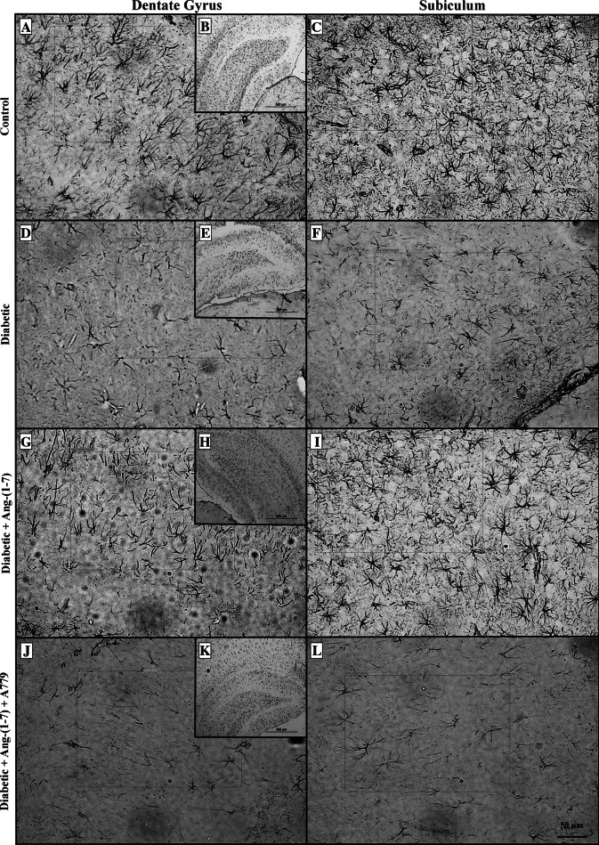
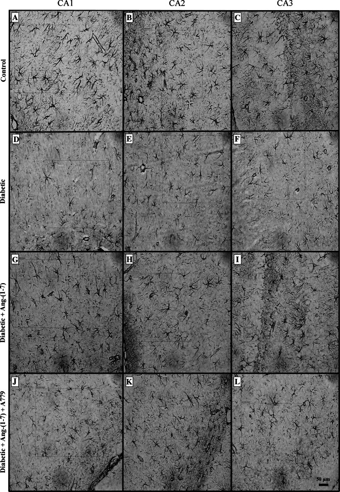
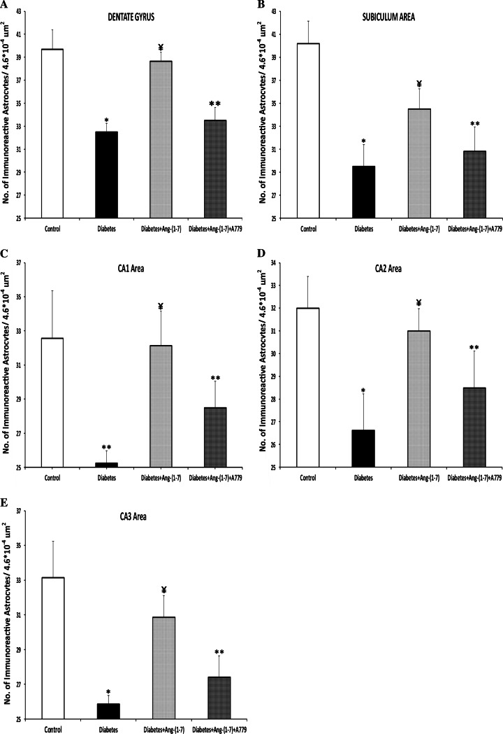
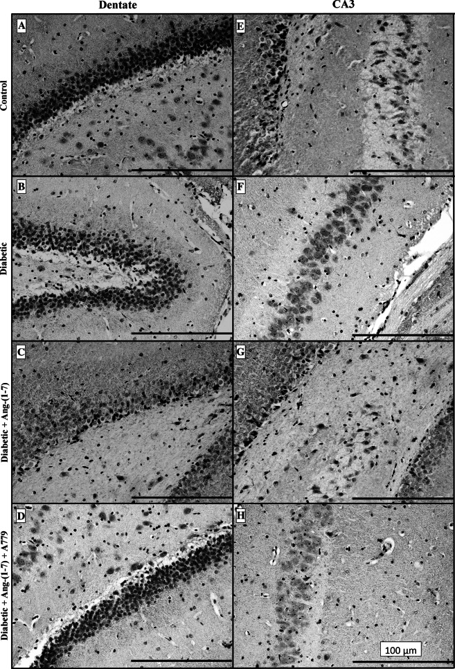
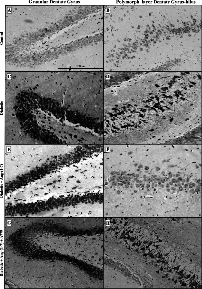
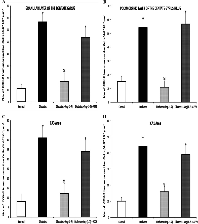
We have previously shown that chronic treatment with angiotensin-(1-7) [Ang-(1-7)] can prevent diabetes-induced cardiovascular dysfunction. However, effect of Ang-(1-7) treatment on diabetes-induced alterations in the CNS is unknown. The aim of this study was to test the hypothesis that treatment with Ang-(1-7) can produce protection against diabetes-induced CNS changes. We examined the effect of Ang-(1-7) on the number of cyclooxygenase-2 (COX-2) immunoreactive neurons and the glial fibrillary acidic protein (GFAP)-immunoreactive astrocytes and assessed the changes in the neuronal growth-associated protein-43 (GAP-43) of the hippocampal formation in streptozotocin-induced diabetes in rats. Animals were sacrificed 30 days after induction of diabetes and/or treatment with Ang-(1-7). Ang-(1-7) treatment significantly prevented diabetes-induced decrease in the number of GFAP immunoreactive astrocytes and GAP-43 positive neurons in all hippocampal regions. Co-administration of A779, a selective Ang-(1-7) receptor antagonist, inhibited Ang-(1-7)-mediated protective effects indicating that Ang-(1-7) produces its effects through activation of receptor Mas. Further, Ang-(1-7) treatment through activation of Mas significantly prevented diabetes-induced increase in the number of the COX-2 immunolabeled neurons in all sub-regions of the hippocampus examined. These results show that Ang-(1-7) has a protective role against diabetes-induced changes in the CNS.


Figures

Fig. 1
Fig. 1
Representative photomicrographs of glial fibrillary acidic protein (GFAP) immunoreactive astrocytes from dentate gyrus (a, d, g, j) and subiculum (c, f, i, l) areas of the hippocampus in control, diabetic, and diabetic + Ang-(1–7) rats and diabetic + Ang-(1–7) + A779. a, b, c Sections showing GFAP immunoreactive astrocytes from control animals; df examples from diabetic group showing visible decrease in the number of GFAP immunoreactive astrocytes, while gi sections from diabetic rats treated with Ang-(1–7) showing a significant increase in the number of GFAP immunoreactive astrocytes. This increase was blocked in diabetic + Ang-(1–7) group when treated with A779 (jl)
Fig. 2
Fig. 2
Representative light photomicrographs of GFAP immunoreactive astrocytes from CA1 (a, d, g, j), CA2 (b, e, h, k), and CA3 (c, f, i, l) areas of the hippocampus in control, diabetic, and diabetic + Ang-(1–7) rats and diabetic + Ang-(1–7) + A779. ac Sections showing GFAP immunoreactive astrocytes from control animals; df examples from diabetic group showing visible decrease in the number of GFAP immunoreactive astrocytes, while gi sections from diabetic rats treated with Ang-(1–7) showing a substantial increase in the number of GFAP immunoreactive astrocytes. This increase was reduced in diabetic + Ang-(1–7) group treated with A779 (jl)
Fig. 3
Fig. 3
Quantitative analysis of the number of GFAP immunoreactive astrocytes in the different areas of the hippocampus: a dentate gyrus, b subiculum, c CA1, d CA2, and e CA3. Diabetic animals showed significant decrease in the number of GFAP immunoreactive astrocytes compared to control group in all the areas of the hippocampus investigated. Treatment of diabetic rats with Ang-(1–7) significantly increased the number of GFAP immunostained astrocytes in all hippocampal areas. This increase was attenuated with in the diabetic animals treated with Ang-(1–7) and A779. Statistical analysis was carried out by one-way ANOVA followed by Bonferroni’s test. Results are expressed are mean ± SEM. *P < 0.001; compared to control and diabetic + Ang-(1–7) groups. **P < 0.001; compared to control group. ¥ P < 0.02; compared to diabetic and diabetic + Ang-(1–7) + A779 groups
Fig. 4
Fig. 4
Representative photomicrographs of GAP-43 immunoreactivity in dentate (ad) and CA3 (eh) areas of the hippocampus from control, diabetic, and diabetic + Ang-(1–7) and diabetic + Ang-(1–7) + A779 rat groups. Dentate and CA3 hippocampal sections from control animals (A and E) showed visible GAP-43 immunoreactive density. The GAP-43 dense labeling was mainly confined to terminals and dendritic layers but few neuronal cell bodies. Hippocampal sections from diabetic rats (b, f) showed prominent GAP-43 immunolabeling density decrease. In contrast, sections from diabetic rats treated with Ang-(1–7) (c, g) showed visible increase of GAP-43 immunolabeling which was blocked in diabetic animals treated with Ang-(1–7) and A779 (d, h)
Fig. 5
Fig. 5
Distribution of COX-2 immunoreactivity in granular dentate gyrus (a, c, e, g) and polymorphic layer of dentate gyrus-hilus (b, d, f, h) areas of the hippocampus in control, diabetic, and diabetic + Ang-(1–7) rats and diabetic + Ang-(1–7) + A779. Diabetes resulted in a substantial increase in COX-2 immunoreactivity in both sub-regions of the dentate hippocampal sector (c, d) compared to faintly labeled control group (a, b). The levels and density of COX-2 immunoreactivity returned to basal levels throughout the dentate hippocampal areas after Ang-(1–7) treatment (e, f). However, marked increase in COX-2 immunoreactivity was observed in diabetic rats treated with Ang-(1–7) and A779 (g, h). Moreover, this marked COX-2 staining was morphologically pyknotic and condensed to a perinuclear location
Fig. 6
Fig. 6
Effect of Ang-(1–7) and Ang-(1–7) + A779 on the COX-2 immunoreactive neurons in the hippocampus of diabetic rats. Quantitative analysis of the number of the COX-2 positive neurons in the granular (a) and polymorphic (b) layers of the dentate gyrus, CA3 (c), and CA1 (d) sub-regions of the rat hippocampus showed a significant increase in all areas of the diabetic animals. Treatment of diabetic rats with Ang-(1–7) resulted in significant reduction in the number of COX-2 immunopositive neurons in all hippocampal sub-regions; while Ang-(1–7) + A779 treatment significantly blocked the decrease in number of the COX-2-immunolabeled cells. Note that there is no significant difference between diabetic-untreated and diabetic Ang-(1–7) + A779-treated groups in the granular and polymorphic layers of the dentate gyrus, CA3 or CA1 areas of the hippocampus. Results are expressed are mean ± SEM. *P < 0.001; compared to control and diabetic + Ang-(1–7) groups. ¥ P < 0.001; compared to diabetic and diabetic + Ang-(1–7) + A779 groups

Similar articles

Cited by

References

    1. Afsari ZH, Renno WM, Abd-El-Basset E (2008) Alteration of glial fibrillary acidic proteins immunoreactivity in astrocytes of the spinal cord diabetic rats. Anat Rec 291:390–399 - PubMed
    1. Al-Maghrebi M, Benter IF, Diz DI (2009) Endogenous angiotensin-(1–7) reduces cardiac ischemia-induced dysfunction in diabetic hypertensive rats. Pharmacol Res 59:263–268 - PMC - PubMed
    1. Artola A, Kamal A, Ramakers GM, Biessels GJ, Gispen WH (2005) Diabetes mellitus concomitantly facilitates the induction of long-term depression and inhibits that of long-term potentiation in hippocampus. Euro J Neurosci 22:169–178 - PubMed
    1. Bagi Z, Erdei N, Papp Z, Edes I, Koller A (2006) Up-regulation of vascular cyclooxygenase-2 in diabetes mellitus. Pharmacol Rep 58:52–56 - PubMed
    1. Barber AJ, Antonetti DA, Gardner TW (2000) Altered expression of retinal occludin and glial fibrillary acidic protein in experimental diabetes. Investig Ophthalmol Vis Sci 41:3561–3568 - PubMed

Publication types

MeSH terms

LinkOut - more resources