. 2012 Dec;33(6):471-83.
doi: 10.1007/s10974-012-9306-9. Epub 2012 Jun 19.

New insights into myosin phosphorylation during cyclic nucleotide-mediated smooth muscle relaxation

Affiliations

New insights into myosin phosphorylation during cyclic nucleotide-mediated smooth muscle relaxation

Sandra Puetz et al. J Muscle Res Cell Motil. 2012 Dec.

Abstract

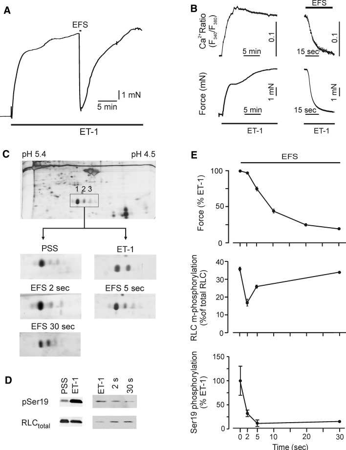
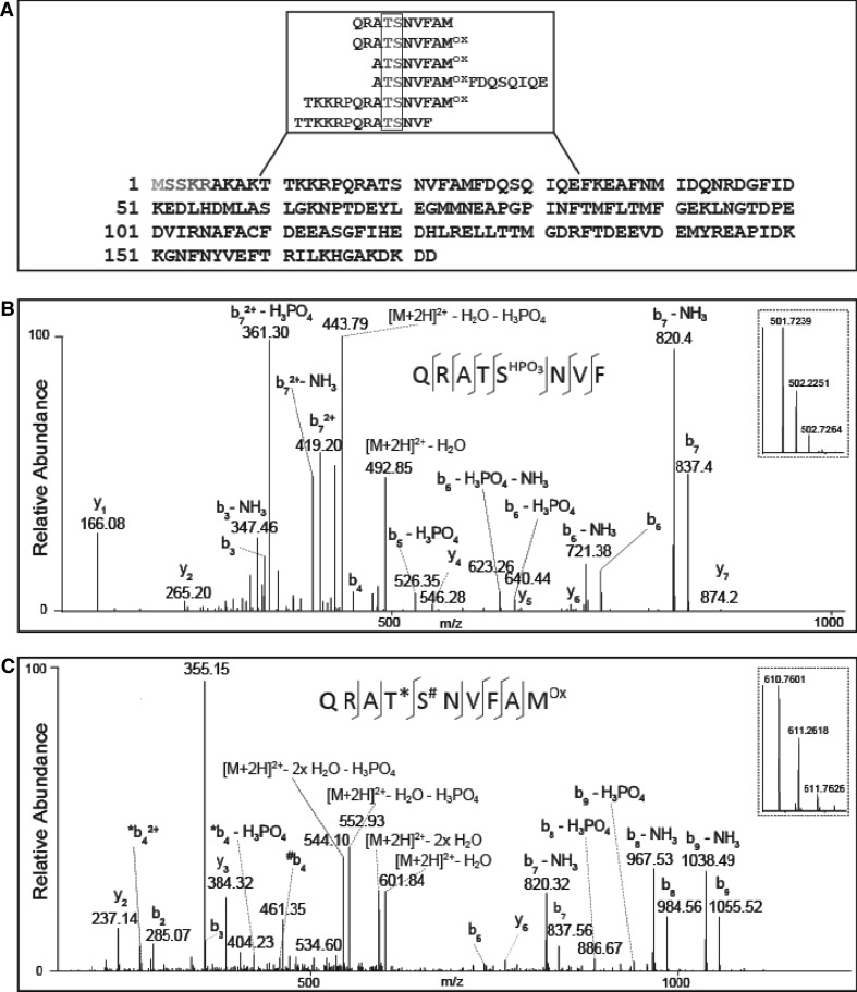
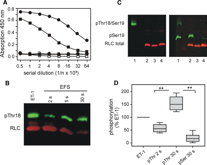
Nitrovasodilators and agonists, via an increase in intracellular cyclic nucleotide levels, can induce smooth muscle relaxation without a concomitant decrease in phosphorylation of the regulatory light chains (RLC) of myosin. However, since cyclic nucleotide-induced relaxation is associated with a decrease in intracellular [Ca(2+)], and hence, a decreased activity of MLCK, we tested the hypothesis that the site responsible for the elevated RLC phosphorylation is not Ser19. Smooth muscle strips from gastric fundus were isometrically contracted with ET-1 which induced an increase in monophosphorylation from 9 ± 1 % under resting conditions (PSS) to 36 ± 1 % determined with 2D-PAGE. Electric field stimulation induced a rapid, largely NO-mediated relaxation with a half time of 8 s, which was associated with an initial decline in RLC phosphorylation to 18 % within 2 s and a rebound to 34 % after 30 s whereas relaxation was sustained. In contrast, phosphorylation of RLC at Ser19 probed with phosphospecific antibodies declined in parallel with force. LC/MS and western blot analysis with phosphospecific antibodies against monophosphorylated Thr18 indicate that Thr18 is significantly monophosphorylated during sustained relaxation. We therefore suggest that (i) monophosphorylation of Thr18 rather than Ser19 is responsible for the phosphorylation rebound during sustained EFS-induced relaxation of mouse gastric fundus, and (ii) that relaxation can be ascribed to dephosphorylation of Ser19, the site considered to be responsible for regulation of smooth muscle tone.


Figures

Fig. 1
Fig. 1
Phosphorylation transients during electrical-field-induced relaxation of endothelin-1 (ET-1) precontracted smooth muscle strips from mouse gastric fundus. a Representative force tracing of ET-1-induced contraction and EFS-induced relaxation (10 Hz, 0.5 ms pulses, duration 30 s) and force recovery. b Simultaneous determination of intracellular Ca2+-transients and force in ET-contracted (left panels) and EFS-relaxed fundus tissues. The ratios of the fluorescence signals excited by 340 nm, and 380 nm (F340/F380) were used as indicator of intracellular [Ca2+]; representative tracings from 6 independent experiments. c Determination of RLC phosphorylation by 2D-PAGE; spot 1 and 2 refer to, respectively, unphosphorylated and monophosphorylated RLC. The intensity of spot 3 amounts to ~14 % of total RLC and does not change significantly in contracted and relaxed preparations. PSS resting conditions prior to ET-1 stimulation, ET-1 plateau of contraction just prior to EFS and 2, 5 and 30 s after starting EFS. d Western blots with pSer19 and RLC total antibody, left panel separation of proteins with PhosTag gels and visualization of immunoreactivity with the odyssey system; right panel separation by 15 % SDS-PAGE and visualization of the immunoreactivity with ECL. e Summary of time courses of EFS-induced relaxation (upper panel), m-phosphorylation of RLC determined by 2D-PAGE (middle panel) and m-phosphorylation of Ser19 normalized to immunoreactive signal obtained with the total RLC antibody and expressed in  % of the ET-1 value (lower panel). A similar result was obtained when pSer19 was expressed relative to the Coomassie stained desmin band. Symbols represent mean ± SEM of 4–9 determinations
Fig. 2
Fig. 2
Identification of RLC and fragment spectra of phosphopeptides. a Sequence of RLC. Amino acid sequences identified by MS are marked in black. In total, a sequence coverage of 97 % was achieved. Phosphorylation of RLC at Thr18/Ser19 (boxed gray letters) was confirmed by multiple peptides (inset). b MS/MS spectrum of the monophosphorylated peptide QRATS*NVF (m/z 501.7239) of myosin regulatory light chain from fully contracted muscle. The phosphorylation site is localized to S19. c MS/MS spectrum of the monophosphorylated peptide QRATSNVFAMOx (m/z 610.7601) of myosin regulatory light chain from muscle treated with EFS for 30 s. The fragment ions observed in the spectrum support monophosphorylation events at T18 (*) as well as S19 (#). The corresponding survey scans for each peptide are displayed as zoom-in views
Fig. 3
Fig. 3
Determination of m-phosphorylation of Thr18 during EFS-induced relaxation using phosphospecific antibodies. a Assessing specificity of anti-phospho-antibody p-MLC (Thr18)-R (Santa Cruz Biotechnology #sc-19848R) by ELISA. As substrate a differently phosphorylated peptide identical to amino acid residues 11-26 of myosin light chain polypeptide 9 Mus musculus (NCBI Protein Data Bank NP_742116) was used. The antibody exhibited a higher affinity against monophosphorylated Thr18 (closed circles) than against diphosphorylation at Ser19 and Thr18 (closed squares). The immunoreactivity against m-phosphorylation at Ser19 (closed diamonds) and the nonphosphorylated peptide (open circles) was very poor and came close to the one measured against ovalbumin (open squares). b Representative western blots with phosphospecific antibodies against m-phosphorylated Thr18, and c pTrh18/pSer19 diphosphorylated RLC (left panel) and m-phosphorylated Ser19 (RLC) and total RLC. The different phosphospecies of RLC were separated by Phos-tag gels, lane 1 mouse tail artery arteries incubated at pCa 6.8 and in the presence of 10 μM microcystin, lane 2–4 gastric fundus contracted with ET-1 (lane 2), relaxed with EFS for 5 s (lane 3) and 30 s (lane 4). d Summary of changes of m-pThr18 during EFS-induced relaxation, pSer19 is replotted from Fig. 1. Bars represent box plots of n = 4 pThr18 and n = 10 pSer19 determinations, ** p < 0.01

Similar articles

Cited by

References

    1. Albrecht K, Schneider A, Liebetrau C, Rüegg JC, Pfitzer G. Exogenous caldesmon promotes relaxation of guinea-pig skinned taenia coli smooth muscles: inhibition of cooperative reattachment of latch bridges? Pflügers Arch. 1997;434:534–542. doi: 10.1007/s004240050433. - DOI - PubMed
    1. Arner A, Pfitzer G. Regulation of cross-bridge cycling by Ca2+ in smooth muscle. Rev Physiol Biochem Pharmacol. 1999;134:63–146. - PubMed
    1. Bárány M, Bárány K. Dissociation of relaxation and myosin light chain dephosphorylation in porcine uterine muscle. Arch Biochem Biophys. 1993;305:202–204. doi: 10.1006/abbi.1993.1412. - DOI - PubMed
    1. Bárány K, Bárány M. Myosin light chains. In: Bárány M, editor. Biochemistry of smooth muscle contraction. San Diego: Academic Press; 1996. pp. 21–35.
    1. Bárány M, Bárány K. Protein phosphorylation during contraction and relaxation. In: Bárány M, editor. Biochemistry of smooth muscle contraction. San Diego: Academic Press; 1996. pp. 321–339.

Publication types

LinkOut - more resources