. 2012 Aug 10;287(33):27417-30.
doi: 10.1074/jbc.M112.360461. Epub 2012 Jun 18.

γ-Aminobutyric acid type A (GABAA) receptor α subunits play a direct role in synaptic versus extrasynaptic targeting

Affiliations

γ-Aminobutyric acid type A (GABAA) receptor α subunits play a direct role in synaptic versus extrasynaptic targeting

Xia Wu et al. J Biol Chem. .

Abstract

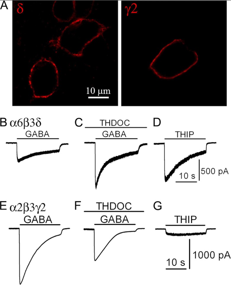
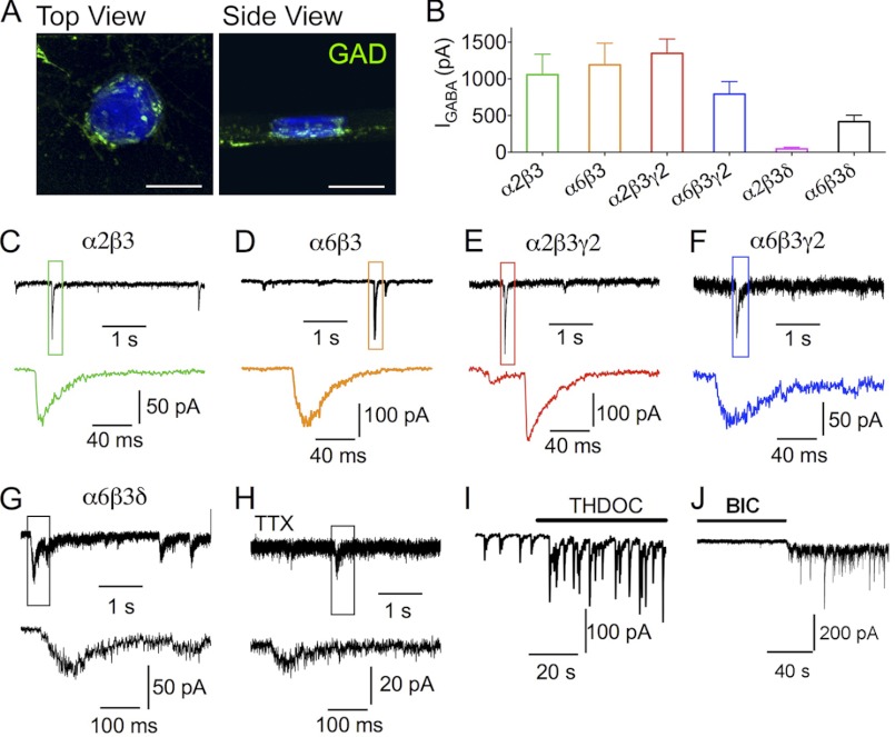
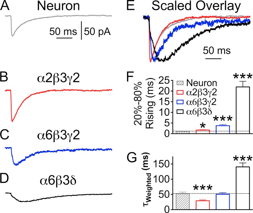
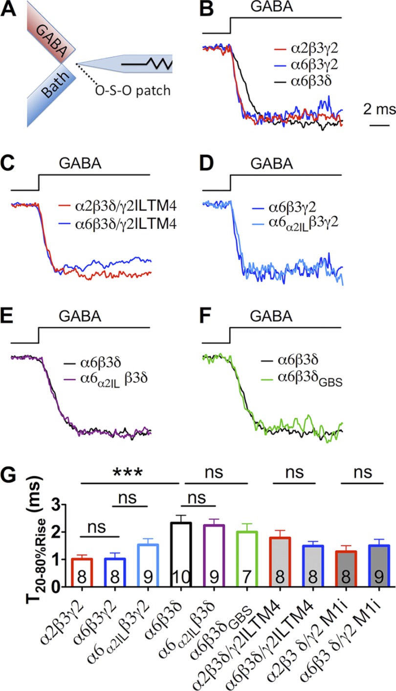
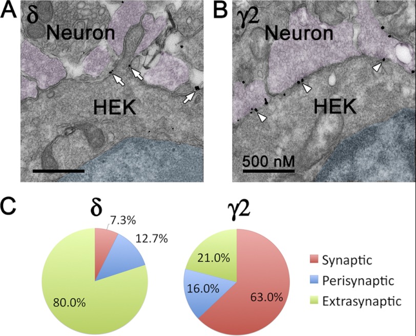
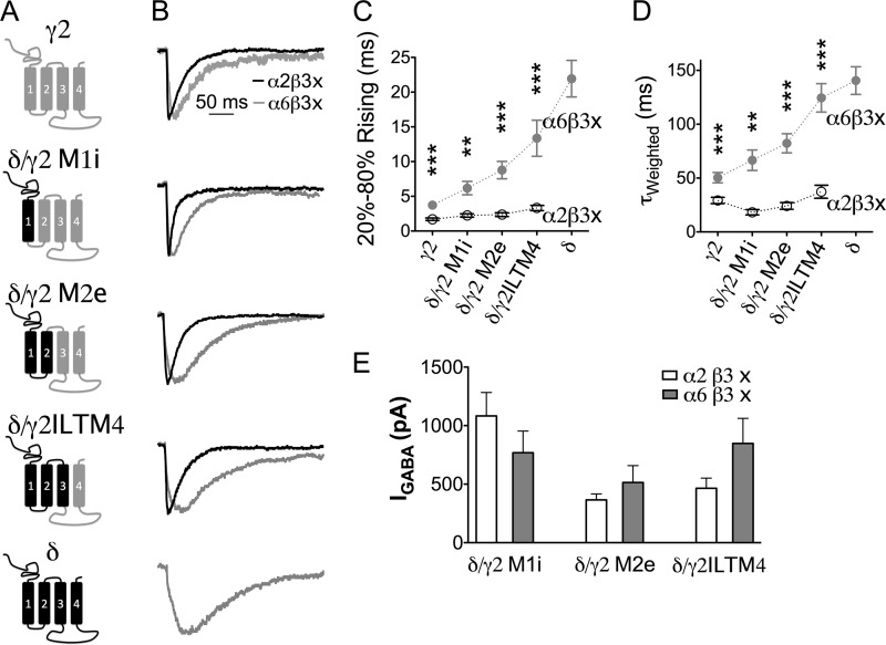
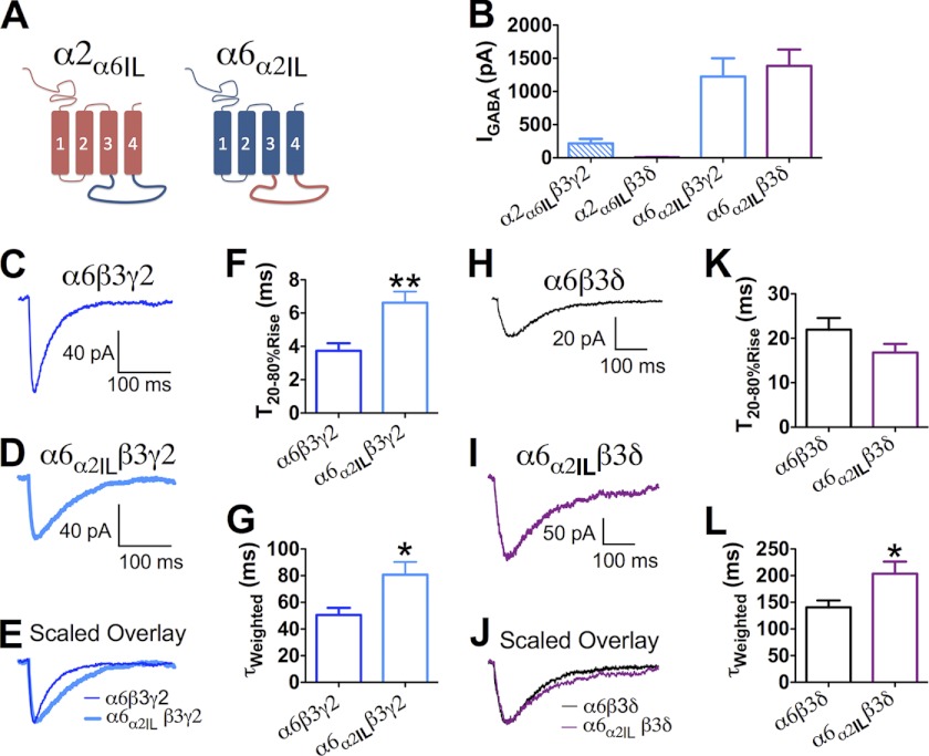
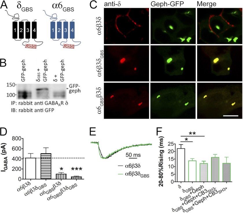
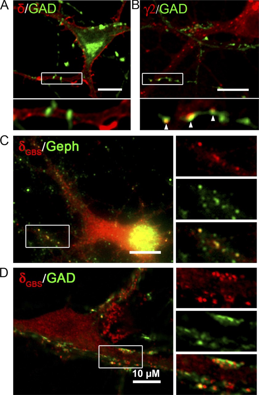
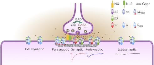
GABA(A) receptors (GABA(A)-Rs) are localized at both synaptic and extrasynaptic sites, mediating phasic and tonic inhibition, respectively. Previous studies suggest an important role of γ2 and δ subunits in synaptic versus extrasynaptic targeting of GABA(A)-Rs. Here, we demonstrate differential function of α2 and α6 subunits in guiding the localization of GABA(A)-Rs. To study the targeting of specific subtypes of GABA(A)-Rs, we used a molecularly engineered GABAergic synapse model to precisely control the GABA(A)-R subunit composition. We found that in neuron-HEK cell heterosynapses, GABAergic events mediated by α2β3γ2 receptors were very fast (rise time ∼2 ms), whereas events mediated by α6β3δ receptors were very slow (rise time ∼20 ms). Such an order of magnitude difference in rise time could not be attributed to the minute differences in receptor kinetics. Interestingly, synaptic events mediated by α6β3 or α6β3γ2 receptors were significantly slower than those mediated by α2β3 or α2β3γ2 receptors, suggesting a differential role of α subunit in receptor targeting. This was confirmed by differential targeting of the same δ-γ2 chimeric subunits to synaptic or extrasynaptic sites, depending on whether it was co-assembled with the α2 or α6 subunit. In addition, insertion of a gephyrin-binding site into the intracellular domain of α6 and δ subunits brought α6β3δ receptors closer to synaptic sites. Therefore, the α subunits, together with the γ2 and δ subunits, play a critical role in governing synaptic versus extrasynaptic targeting of GABA(A)-Rs, possibly through differential interactions with gephyrin.


Figures

FIGURE 1.
FIGURE 1.
Recombinant GABAA-Rs with distinct pharmacological properties. A, comparable expression level of α2β3γ2 and α6β3δ receptors on HEK cell membranes, revealed by surface staining without permeabilization. B–D, pharmacological responses of HEK 293T cells co-expressing α6, β3, and δ subunits. B, representative trace showing the whole-cell GABA (100 μm) current in a HEK 293T cell transfected with α6β3δ. C, positive modulation of GABA-induced current by a neurosteroid THDOC (100 nm). D, THIP (100 μm) acts as a super-agonist on α6β3δ GABAA-Rs. E–G, pharmacological responses of HEK 293T cells co-expressing α2, β3, and γ2 subunits. E, representative trace showing whole-cell GABA (100 μm) current in a α2β3γ2-transfected HEK 293T cells. F, GABA induces a smaller whole-cell current in the presence of THDOC (100 nm). G, THIP acts as a partial agonist for the α2β3γ2-GABAA-Rs.
FIGURE 2.
FIGURE 2.
GABAA receptors with distinct subunit combinations mediate IPSCs in HEK cells. A, three-dimensional reconstitution of Z-stack confocal images showing the GABAergic nerve terminals (green) on the surface of an NL2-transfected HEK cell (blue). Scale bar, 20 μm. B, whole-cell currents (means ± S.E.) induced by 100 μm GABA in HEK cells expressing α2β3, α6β3, α2β3γ2, α6β3γ2, α2β3δ, and α6β3δ receptors. C–G, representative traces showing the IPSCs recorded in HEK cells co-expressing NL2 with α2β3 (C), α6β3 (D), α2β3γ2 (E), α6β3γ2 (F), or α6β3δ (G) GABAA receptors. H, miniature IPSCs recorded from a HEK cell expressing NL2 and α6β3δ receptors in the presence of tetrodotoxin (TTX) (0.5 μm). Lower panels show the expanded views of the boxed IPSCs from the top traces. I, THDOC (100 nm) increases the amplitude of IPSCs in HEK cells co-expressing α6β3δ and NL2. J, application of bicuculline (BIC) (20 μm) reduces the base-line current and the noise level, revealing the tonic current in HEK cells expressing NL2 and α6β3δ receptors.
FIGURE 3.
FIGURE 3.
Quantitative analysis of the kinetics of IPSCs recorded from HEK 293T cells transfected with NL2 and different sets of GABAA-R subunits. A, average trace of IPSCs in a neuron. B–D, average traces of IPSCs mediated by α2β3γ2 (B), α6β3γ2 (C), or α6β3δ (D) receptors. E, scaled overlay of IPSCs from A–D, showing the difference in rising and decay phases. F and G, pooled kinetics data of the IPSCs recorded from neurons and HEK cells expressing α2β3γ2, α6β3γ2, and α6β3δ receptors. F, 20–80% rising time, and G, weighted time constant (τweighted) of sIPSCs. *, p < 0.05; ***, p < 0.001 (one-way ANOVA followed by Bonferroni's pairwise comparison).
FIGURE 4.
FIGURE 4.
Onset kinetics of recombinant GABAA-Rs. A, schematic diagram illustrating the fast drug application system. Fast GABA application was achieve by starting the GABA perfusion and stopping the flow of bath solution instantaneously. B–F, representative traces of GABA-induced currents on outside-out patches excised from transfected HEK cells. B, rise time of α6β3δ-Rs was slower than that of α2β3γ2- and α6β3γ2-Rs. C, no significant difference in the onset kinetics when substituting the α2 with α6 subunit in δ/γ2IL-TM4 chimeric receptors. D and E, insertion of α2IL into the α6 subunit (α6α2IL) did not change the onset kinetics of α6-containing receptors. F, insertion of the GBS into the δ subunit (δGBS) did not change the onset kinetics of the α6β3δ receptors. G, polled data showing the 20–80% rise time of each recombinant GABAA-R. ***, p < 0.001 (one-way ANOVA followed by Bonferroni's pairwise comparison). ns, no significance.
FIGURE 5.
FIGURE 5.
Ultrastructural localization of α6β3δ- and α2β3γ2-GABAA-Rs in the hetero-reconstituted synapses. A and B, transfected HEK cells in close contact with nerve terminals. The nuclei of HEK cells are shaded cyan. Nerve terminals containing synaptic vesicles are shown in magenta. A, α6β3δ receptors were predominantly localized at extrasynaptic or perisynaptic membranes. Arrows, silver-enhanced gold particles labeling the δ subunit-containing receptors. B, α2β3γ2 receptors were mainly localized at synaptic sites. Arrowheads, immunogold labeling of the GFP-γ2 subunit. C, pie graphs showing the percentage of synaptic, perisynaptic, and extrasynaptic labeling in HEK cells expressing α6β3δ or α2β3γ2 receptors.
FIGURE 6.
FIGURE 6.
Different α subunits contribute to the distinct kinetics of sIPSCs. A, schematic representation of a series of δ/γ2 chimeric subunits. IL, intracellular loop; TM, transmembrane domain. B, average traces of sIPSCs recorded from representative HEK cells expressing γ2 subunits, δ subunits, or δ/γ2 chimeras together with either α2 and β3 or α6 and β3 subunits and NL2 “x” in a2b3x or a6b3x stands for either g2, d, or their chimeric subunit. C, pooled data (mean ± S.E.) showing the 20–80% rise time of IPSCs in HEK cells expressing different subunit combinations. Each chimera showed significantly faster rise phase when paired up with the α2 subunit, compared with when the α6 subunit was their assembly partner. **, p < 0.01; ***, p < 0.001, two-tailed t test. D, average weighted time constant (τweighted) of sIPSCs mediated by different subunit combinations (mean ± S.E.). E, GABA-induced current (100 μm) mediated by δ/γ2 chimera-containing receptors in HEK cells.
FIGURE 7.
FIGURE 7.
α2 subunit intracellular domain was not sufficient to determine synaptic receptor targeting. A, schematic diagram indicating the structure of α2α6IL and α6α2IL chimeras. B, whole-cell current induced by 100 μm GABA in HEK cells expressing α2α6ILβ3γ2, α2α6ILβ3δ, α6α2ILβ3γ2, and α6α2ILβ3δ receptors. C and D, averaged sIPSC traces recorded from HEK cells expressing α6β3γ2 or α6α2ILβ3γ2 receptors. E, scaled overlay of α6β3γ2 or α6α2ILβ3γ2 receptor-mediated IPSCs. F and G, pooled data (mean ± S.E.) showing the comparison of the 20–80% rising and τweighted of α6β3γ2 and α6α2ILβ3γ2 receptor-mediated IPSCs. H and I, representative traces showing the averaged sIPSC events from HEK cells expressing α6β3δ (H) and α6α2ILβ3δ (I) receptors. J, scaled overlay of α6β3δ or α6α2ILβ3δ receptor-mediated IPSCs. K and L, pooled data comparing the 20–80% rising and τweighted of α6β3δ and α6α2ILβ3δ receptor-mediated IPSCs. *, p < 0.05; **, p < 0.01
FIGURE 8.
FIGURE 8.
Interaction with gephyrin targets α6β3δ-containing GABAA-Rs to synaptic sites of reconstituted synapses. A, schematic representation of the δGBS and α6GBS chimeras. B, GFP-gephyrin (geph) was co-precipitated with the δGBS chimera. C, α6β3δGBS and α6GBSβ3δ receptors were found to co-localize with gephyrin-GFP in big intracellular aggregates when co-expressed in HEK cells, demonstrating the interaction between δGBS and α6GBS subunits and gephyrin; α6β3δ receptors showed no co-localization with gephyrin-GFP. Scale bar, 10 μm. D, whole-cell GABA current in HEK cells expressing the δGBS and/or α6GBS chimeras. E, sample traces of sIPSCs mediated by α6β3δ or α6β3δGBS receptors. The events were scaled to the same amplitude and aligned according to the initial rise time. F, pooled data of sIPSC rise time in different groups. The α6β3δGBS receptor-mediated sIPSCs showed a rise phase significantly faster than that of α6β3δ receptors. Co-expression of gephyrin or gephyrin plus collybistin did not further change the IPSC rise time. *, p < 0.05; **, p < 0.01; ***, p < 0.001 (one-way ANOVA followed by Bonferroni's pairwise comparison).
FIGURE 9.
FIGURE 9.
Incorporation of the gephyrin-binding site induces clustering of α6β3δ-receptors at postsynaptic sites in neurons. A, hypothalamic neurons co-transfected with α6, β3, and δ subunits were double immunolabeled for the δ subunit and GAD. Immunoreactivity for the δ subunit was diffusely localized on the neuronal surface. B, neurons were co-transfected with α2, β3, and mycγ2 subunits, followed by GAD and surface mycγ2 double staining. The γ2 subunit-containing receptors formed puncta along the dendrites, some of which were apposed to GAD puncta. C and D, neurons transfected with α6GBS, β3, and δGBS subunits were double immunolabeled for the δGBS subunit and gephyrin (Geph) (C) or δGBS and GAD (D). The δGBS subunit-containing receptors formed clusters in neurons. A portion of the δGBS subunit-containing receptors were co-localized with gephyrin clusters (right panels in C) or with GAD puncta (right panels in D), suggesting a synaptic localization.
FIGURE 10.
FIGURE 10.
Model for synaptic versus extrasynaptic GABAA-R targeting. The α2β3 and α2β3γ2 GABAA-Rs are clustered at synaptic sites, mediating fast IPSCs. The α6β3δ GABAA-Rs are localized at extrasynaptic sites, mediating very slow IPSCs, which may be partly due to a lack of binding with gephyrin (Geph). Importantly, the α6β3 and α6β3γ2 GABAA-Rs are likely localized at perisynaptic sites, resulting in IPSC kinetics in-between that of α2β3γ2 and α6β3δ GABAA-Rs. The α6GBSβ3δGBS GABAA-Rs are brought closer to synaptic sites.

References

    1. Luscher B., Fuchs T., Kilpatrick C. L. (2011) GABAA receptor trafficking-mediated plasticity of inhibitory synapses. Neuron 70, 385–409 - PMC - PubMed
    1. Jacob T. C., Moss S. J., Jurd R. (2008) GABAA receptor trafficking and its role in the dynamic modulation of neuronal inhibition. Nat. Rev. Neurosci. 9, 331–343 - PMC - PubMed
    1. Olsen R. W., Sieghart W. (2009) GABAA receptors. Subtypes provide diversity of function and pharmacology. Neuropharmacology 56, 141–148 - PMC - PubMed
    1. Sigel E., Baur R., Boulineau N., Minier F. (2006) Impact of subunit positioning on GABAA receptor function. Biochem. Soc. Trans. 34, 868–871 - PubMed
    1. Farrant M., Nusser Z. (2005) Variations on an inhibitory theme. Phasic and tonic activation of GABAA receptors. Nat. Rev. Neurosci. 6, 215–229 - PubMed

Publication types

LinkOut - more resources