. 2012 Dec;56(6):2375-86.
doi: 10.1002/hep.25900.

The liver-specific tumor suppressor STAT5 controls expression of the reactive oxygen species-generating enzyme NOX4 and the proapoptotic proteins PUMA and BIM in mice

Affiliations

The liver-specific tumor suppressor STAT5 controls expression of the reactive oxygen species-generating enzyme NOX4 and the proapoptotic proteins PUMA and BIM in mice

Ji Hoon Yu et al. Hepatology. 2012 Dec.

Abstract

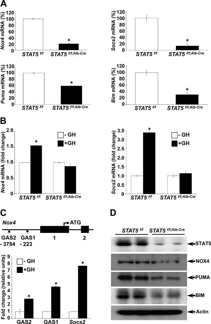
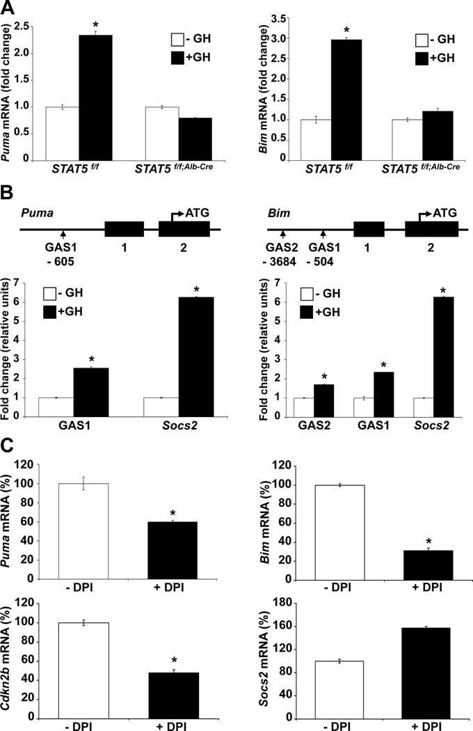
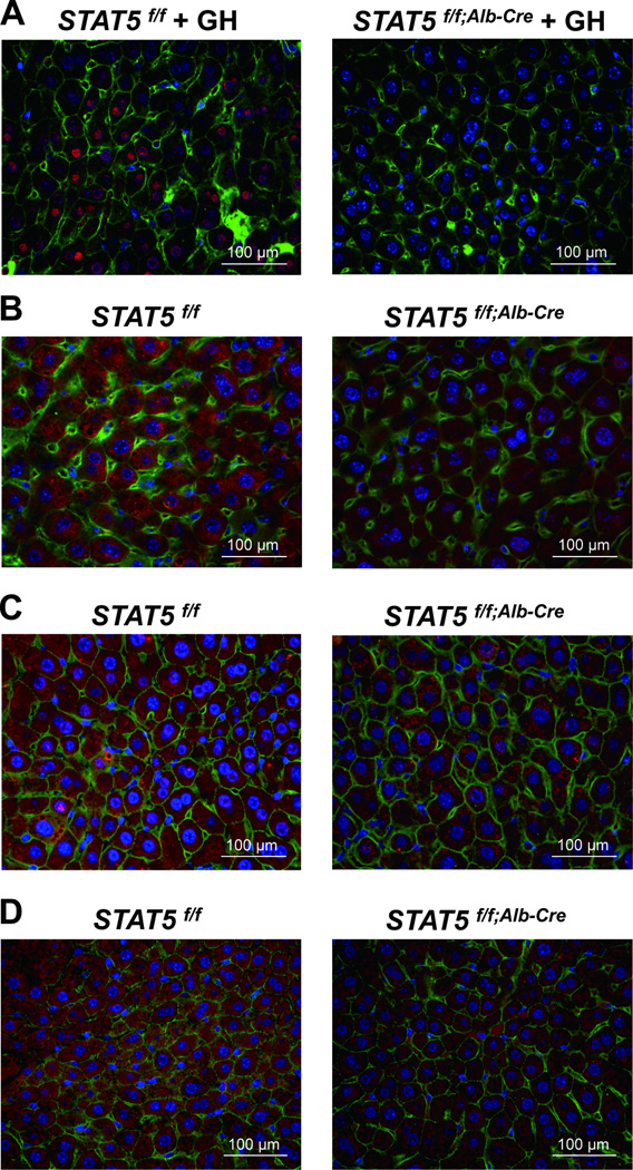
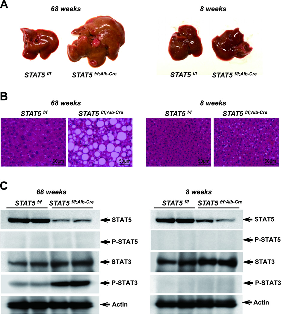
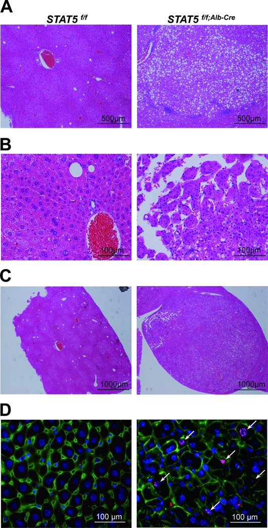
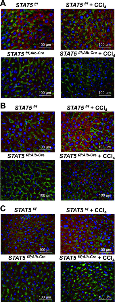
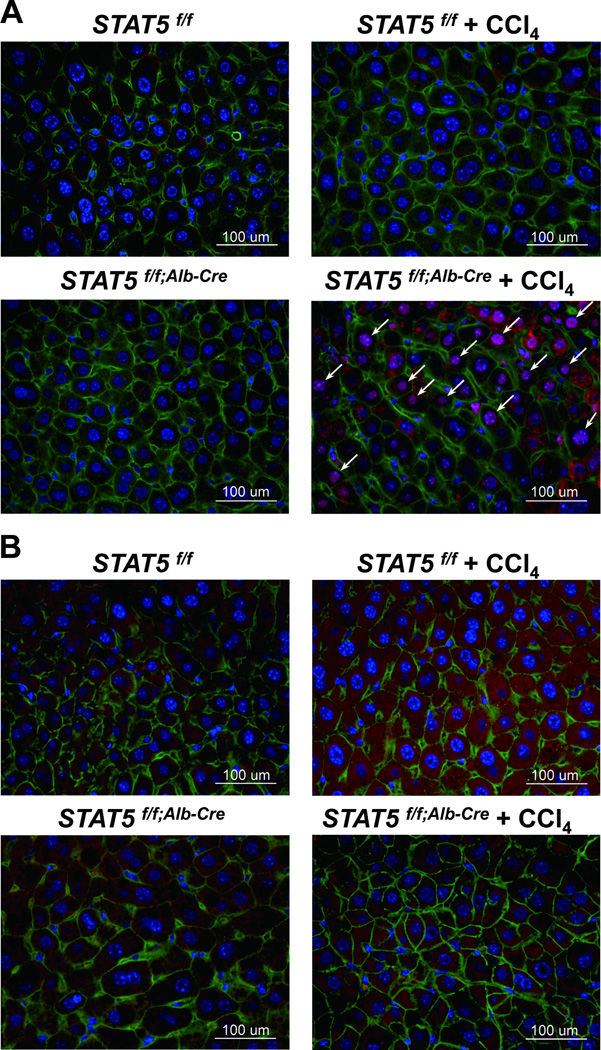
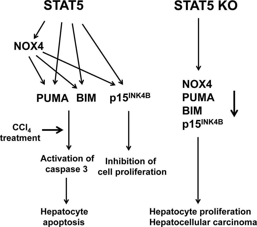
Loss of signal transducer and activator of transcription 5 (STAT5) from liver tissue results in steatosis and enhanced cell proliferation. This study demonstrates that liver-specific Stat5-null mice develop severe hepatic steatosis as well as hepatocellular carcinomas at 17 months of age, even in the absence of chemical insults. To understand STAT5's role as a tumor suppressor, we identified and investigated new STAT5 target genes. Expression of Nox4, the gene encoding the reactive oxygen species (ROS)-generating enzyme NOX4, was induced by growth hormone through STAT5. In addition, the genes encoding the proapoptotic proteins PUMA and BIM were induced by growth hormone through STAT5, which bound to GAS motifs in the promoter regions of all three genes. We further show that STAT5-induced activation of Puma and Bim was dependent on NOX4. Treatment of mice with transforming growth factor-β, an inducer of apoptosis, resulted in cleaved caspase-3 in control but not in liver-specific Stat5-null mice. This study demonstrates for the first time that cytokines through STAT5 regulate the expression of the ROS-generating enzyme NOX4 and key proapoptotic proteins.

Conclusion: STAT5 harnesses several distinct signaling pathways in the liver and thereby functions as a tumor suppressor. Besides suppressing the activation of STAT3, STAT5 induces the expression of proapoptotic genes and the production of ROS.


Figures

Figure 1
Figure 1
STAT5 regulates Nox4 expression through STAT5 binding to conserved GAS sites in the Nox4 gene promoter in liver. (A) Expression of Nox4, Puma, Bim and Socs2 was analyzed by quantitative real-time PCR in liver tissue from Stat5f/f and Stat5f/f;Alb-Cre mice. Values are shown as means ± SD. (B) mRNA expression of Nox4 and Socs2 in Stat5f/f and Stat5f/f;Alb-Cre mice. Mice were injected with GH, tissue was harvested after four hours and RNA was analyzed by quantitative real-time PCR. All values represent means ± SD. (C) Schematic of the Nox4 gene. Vertical boxes indicate exons and conserved GAS sequences are marked. Chromatin immunoprecipitation (ChIP) analysis of STAT5 binding to the putative GAS sites. Stat5f/f mice were injected with GH, tissue was harvested after 45 minutes and binding to GAS sites was analyzed by quantitative real-time PCR. DNA was amplified from STAT5-precipitated complexes using specific primers spanning GAS motifs in the Socs2 and Nox4 genes. All values represent means ± SD from 3 independent experiments performed in triplicates. (D) Levels of NOX4, PUMA and BIM in liver tissue from Stat5f/f and Stat5f/f;Alb-Cre mice. Expression of NOX4, PUMA and BIM was determined by western blotting. *P < .05; compared with corresponding controls.
Figure 2
Figure 2
STAT5 regulates expression of Puma and Bim through STAT5 binding to GAS sites in the Puma and Bim gene promoters in liver. (A) mRNA expression of Puma and Bim in Stat5f/f and Stat5f/f;Alb-Cre mice injected with GH. Mice were injected with GH and tissue was harvested after 4 hours. Expression of Puma and Bim was analyzed by quantitative real-time PCR in liver tissue from Stat5f/f and Stat5f/f;Alb-Cre mice. All values represent means ± SD. (B) Schematic of the Puma and Bim genes. Vertical boxes indicate exons and the location of conserved GAS sequences is shown. Chromatin immunoprecipitation (ChIP) analysis of STAT5 binding to the putative GAS sites. Stat5f/f mice were treated with GH and tissue was harvested after 45 minutes. Binding to GAS sites was analyzed by quantitative real-time PCR. DNA was amplified from STAT5-precipitated complexes using specific primers for known (Socs2) and suspected (Puma and Bim) GAS regions. All values represent means ± SD from 3 independent experiments performed in triplicates. (C) mRNA expression of Puma, Bim, Cdkn2b and Socs2 in immortalized wildtype hepatocyte of murine origin. The cells were treated with DPI for 2 hours. Expression of Puma, Bim, Cdkn2b and Socs2 mRNA was analyzed by quantitative real-time PCR. All values represent means ± SD from 3 independent experiments. *P < .05; compared with corresponding controls.
Figure 3
Figure 3
Immunostaining of phospho-STAT5, NOX4, PUMA and BIM in Stat5f/f and Stat5f/f;Alb-Cre mice. (A) Livers from Stat5f/f and Stat5f/f;Alb-Cre mice were harvested after GH injection and analyzed for p-STAT5 expression using immunofluorescence staining with anti-p-STAT5 (red), anti-β-catenin (green) antibodies and DAPI (blue). (B–D) Livers from Stat5f/f and Stat5f/f;Alb-Cre mice were harvested and analyzed for NOX4 (B), PUMA (C) and BIM (D) expression using immunofluorescence staining with anti-NOX4 (red), anti-PUMA (red), anti-BIM (red), anti-β-catenin (green) antibodies and DAPI (blue).
Figure 4
Figure 4
Loss of STAT5 induces development of tumor in Stat5f/f;Alb-Cre mice. (A) Liver of Stat5f/f and Stat5f/f;Alb-Cre mice at 17 months (left) and 2 months (right). (B) Hematoxylin and eosin (H&E) staining of liver sections from Stat5f/f and Stat5f/f;Alb-Cre mice. (C) Level of STAT5, p-STAT5, STAT3 and p-STAT3 in liver tissues from Stat5f/f and Stat5f/f;Alb-Cre mice. Expression of STAT5, p-STAT5, STAT3 and p-STAT3 was determined by western blotting.
Figure 5
Figure 5
Histological analyses and immunostaining of phospho-Histone H3 in liver tissue from Stat5f/f and Stat5f/f;Alb-Cre mice. (A–C) Hematoxylin and eosin (H&E) staining of liver sections from Stat5f/f and Stat5f/f;Alb-Cre mice at 17 months of age. Hepatosteatosis (A) and hepatocellular carcinoma (HCC) (B) were only observed in liver-specific Stat5-null mice. Nodules were also observed only in liver-specific Stat5-null mice (C). (D) Liver tissue from 17 months old Stat5f/f and Stat5f/f;Alb-Cre mice was harvested and analyzed for phospho-Histone H3 using immunofluorescence staining with anti-phospho-Histone H3 (red), anti-β-catenin (green) antibodies and DAPI (blue).
Figure 6
Figure 6
Immunostaining of NOX4, PUMA and BIM in Stat5f/f and Stat5f/f;Alb-Cre mice injected with CCl4. (A–C) Liver tissue from Stat5f/f and Stat5f/f;Alb-Cre mice was harvested after 12 weeks of CCl4 injection and analyzed for expression of NOX4 (A), PUMA (B) and BIM (C) using immunofluorescence staining with anti-NOX4 (red), anti-PUMA (red), anti-BIM (red), anti-β-catenin (green) antibodies and DAPI (blue).
Figure 7
Figure 7
Immunostaining of Ki-67 and cleaved caspase-3 in Stat5f/f and Stat5f/f;Alb-Cre mice injected with CCl4. (A) Liver tissue from Stat5f/f and Stat5f/f;Alb-Cre mice was harvested after 12 weeks of CCl4 injection and analyzed for Ki-67 using immunofluorescence staining with anti-Ki-67 (red), anti-β-catenin (green) antibodies and DAPI (blue). (B) Liver tissue from Stat5f/f and Stat5f/f;Alb-Cre mice was harvested after 12 weeks of CCl4 injection and analyzed for cleaved caspase-3 using immunofluorescence staining with anti-cleaved caspase-3 (red), anti-β-catenin (green) antibodies and DAPI (blue).
Figure 8
Figure 8
Proposed model of STAT5-regulated apoptosis in hepatocytes. Previous studies have shown that liver-specific STAT5-null mice develop hepatosteatosis and HCC upon treatment with CCl4. STAT5 regulates key cell cycle inhibitor and apoptotic genes. STAT5 directly activates the genes encoding NOX4, PUMA and BIM and the cell cycle inhibitor p15INK4B. NOX4 can also control PUMA, BIM and p15INK4B. We propose that loss of STAT5 induces hepatocyte proliferation and HCC upon CCl4 challenge as a result of decreased levels of PUMA, BIM and p15INK4B.

References

    1. Hennighausen L, Robinson GW. Interpretation of cytokine signaling through the transcription factors STAT5A and STAT5B. Genes Dev. 2008;22:711–721. - PMC - PubMed
    1. Zhang Q, Wang HY, Liu X, Wasik MA. STAT5A is epigenetically silenced by the tyrosine kinase NPM1-ALK and acts as a tumor suppressor by reciprocally inhibiting NPM1-ALK expression. Nat Med. 2007;13:1341–1348. - PubMed
    1. Hosui A, Kimura A, Yamaji D, Zhu BM, Na R, Hennighausen L. Loss of STAT5 causes liver fibrosis and cancer development through increased TGF-beta and STAT3 activation. J Exp Med. 2009;206:819–831. - PMC - PubMed
    1. Mallette FA, Gaumont-Leclerc MF, Ferbeyre G. The DNA damage signaling pathway is a critical mediator of oncogene-induced senescence. Genes Dev. 2007;21:43–48. - PMC - PubMed
    1. Calabrese V, Mallette FA, Deschenes-Simard X, Ramanathan S, Gagnon J, Moores A, et al. SOCS1 links cytokine signaling to p53 and senescence. Mol Cell. 2009;36:754–767. - PubMed

Publication types

MeSH terms