Comparative Study
. 2012 Jul;14(7):931-41.
doi: 10.1093/neuonc/nos125. Epub 2012 Jun 18.

Clinical and molecular characteristics of congenital glioblastoma

Affiliations
Comparative Study

Clinical and molecular characteristics of congenital glioblastoma

Margaret E Macy et al. Neuro Oncol. 2012 Jul.

Abstract

Congenital glioblastoma (cGBM) is an uncommon tumor of infancy with a reported variable but often poor cure rate, even with intensive therapy. Five patients with cGBMs, arising de novo and not in familial tumor predisposition kindreds, were studied for histological and biological features, using Affymetrix microarray. Tumors were large, often associated with hemorrhage, extended into the thalamus, and often bulged into the ventricles. One patient died acutely from bleeding at the time of operation. The 4 surviving patients underwent surgery (1 gross total resection, 3 subtotal resections or biopsies) and moderate intensity chemotherapy without radiation, and remain progression-free at a median time of 36 months (range, 30-110 months). Affymetrix microarrays measured gene expression on the 3 cGBMs from which frozen tissue was available. Unsupervised hierarchical clustering of cGBMs versus 168 other central nervous system tumors demonstrated that cGBMs clustered most closely with other high-grade gliomas. Gene expression profiles of cGBMs were compared with non-congenital pediatric and adult GBMs. cGBMs demonstrated marked similarity to both pediatric and adult GBMs, with only 31 differentially expressed genes identified (false discovery rate, <0.05). Unique molecular features of cGBMs included over-expression of multiple genes involved in glucose metabolism and tissue hypoxia. cGBMs show histological and biological overlap with pediatric and adult GBMs but appear to have a more favorable outcome, with good response to moderate intensity chemotherapy with only subtotal resection or biopsy. Further study may determine whether identified gene expression differences contribute to the improved survival seen in these tumors.


Figures
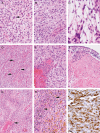
Fig. 1.
Fig. 1.
Histopathological characteristics of congenital GBMs (cGBMs). (A) Tumors were moderately hypercellular glial tumors with a relatively monotonous tumor cell population; note mitotic activity (arrow). Hematoxylin and eosin (H&E), 600×. (B) A second cGBM manifesting a more spindled appearance, but, again, relative monotony in terms of cell size and shape. H&E, 400×. (C) Squash preparation highlights the absence of significant pleomorphism or prominent nucleoli in the tumor cells. Note the delicate fibrillar eosinophilic cytoplasm in the cells. H&E, 600×. (D) Most tumors manifested increased vascular density, sometimes consisting of delicate, arcuate vasculature without thrombosis or microvascular proliferation (arrows). H&E, 200×. (E) Two of the cGBMs showed focal microvascular proliferation and vascular thromboses. H&E, 400×. (F) Tumors were deep-seated and located adjacent to ventricle, abutting the ependymal lining (top), and this example also demonstrates extensive zonal necrosis (bottom). H&E, 400×. (G) Pseudopalisading necrosis and vascular thrombosis (arrow) were identified in several cGBMs. H&E, 200×. (H) Hemosiderin pigment deposition (lower right) was extensive in one case, indicating subacute to chronic bleeding into tumor. This case also manifested Gamna-Gandy body formation (arrows), verifying chronic repetitive bleeding into the tumor. H&E, 400×. (I) Immunoreactivity for glial fibrillary acidic protein (GFAP) was present in all cases. Immunostaining for GFAP with light hematoxylin counterstain, 600×.
Fig. 2.
Fig. 2.
Hierarchical clustering of central nervous system (CNS) tumors. Microarray data from 171 CNS tumors, including a variety of pediatric primary CNS tumors as well as radiation-induced glioblastomas (GBMs) and 6 adult GBMs, were used as input into an agglomerative clustering algorithm. Genes with expression or variability in the bottom 25% across all samples were omitted due to their non-informative nature. The majority of gliomas clustered under one major branch, shown here. The three congenital GBMs for which microarray data were available are marked with arrows. H, high-grade glioma; L, low-grade glioma; C, congenital GBM; E, ependymoma; N, mixed glioneuronal.
Fig. 3.
Fig. 3.
Gene expression of epidermal growth factor receptor (EGFR) and platelet-derived growth factor receptor α (PDGFRα). (A) Gene expression, as measured by Affymetrix U133 Plus2 GeneChip microarray, is shown for adult, pediatric and congenital glioblastomas (GBMs) for EGFR. (B) Similarly, gene expression for adult, pediatric and congenital GBMs for PDGFRα. Data was normalized using the GC robust multi-array average algorithm. Gene expression is shown as absolute values on a linear scale. Abbreviations: aGBM, adult glioblastoma; cGBM, congenital glioblastoma; pGBM, pediatric glioblastoma.
Fig. 4.
Fig. 4.
Principal components analysis (PCA) of congenital, pediatric, and adult GBMs. PCA was performed using genes which distinguish mesenchymal, proliferative and proneural subtypes in adult GBMs. Pediatric and adult GBMs were assigned the label of the subtype with the highest average across all Z-scores for genes that distinguished that subtype. cGBM, congenital glioblastoma.

References

    1. Severino M, Schwartz ES, Thurnher MM, Rydland J, Nikas I, Rossi A. Congenital tumors of the central nervous system. Neuroradiology. 2010;52(6):531–548. - PubMed
    1. Mazewski CM, Hudgins RJ, Reisner A, Geyer JR. Neonatal brain tumors: a review. Semin Perinatol. 1999;23(4):286–298. - PubMed
    1. Rickert CH, Probst-Cousin S, Gullotta F. Primary intracranial neoplasms of infancy and early childhood. Childs Nerv Syst. 1997;13(10):507–513. - PubMed
    1. Jellinger K, Sunder-Plassmann M. Connatal intracranial tumours. Neuropadiatrie. 1973;4(1):46–63. - PubMed
    1. Manoranjan B, Provias JP. Congenital brain tumors: diagnostic pitfalls and therapeutic interventions. J Child Neurol. 2011;26(5):599–614. - PubMed

Publication types

MeSH terms