. 2012 Jul 3;109(27):11025-30.
doi: 10.1073/pnas.1206555109. Epub 2012 Jun 18.

Purified and synthetic Alzheimer's amyloid beta (Aβ) prions

Affiliations

Purified and synthetic Alzheimer's amyloid beta (Aβ) prions

Jan Stöhr et al. Proc Natl Acad Sci U S A. .

Abstract

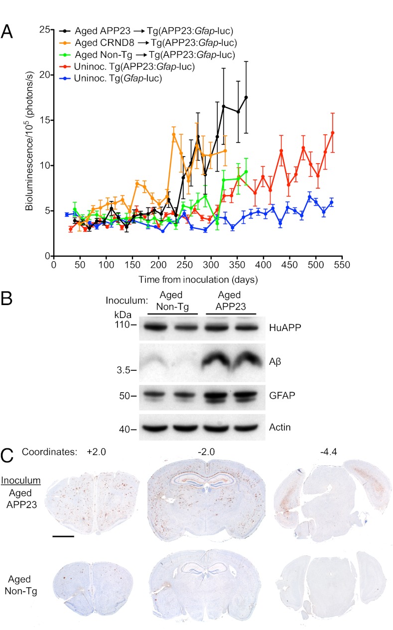
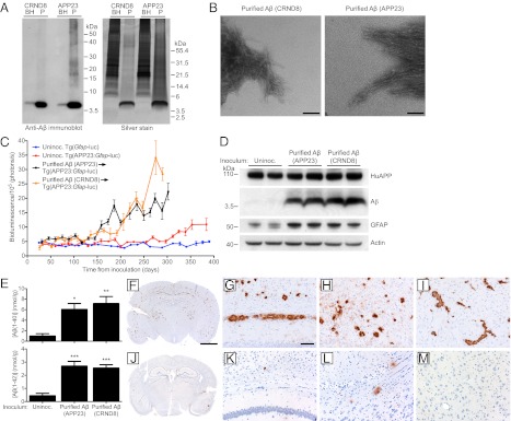
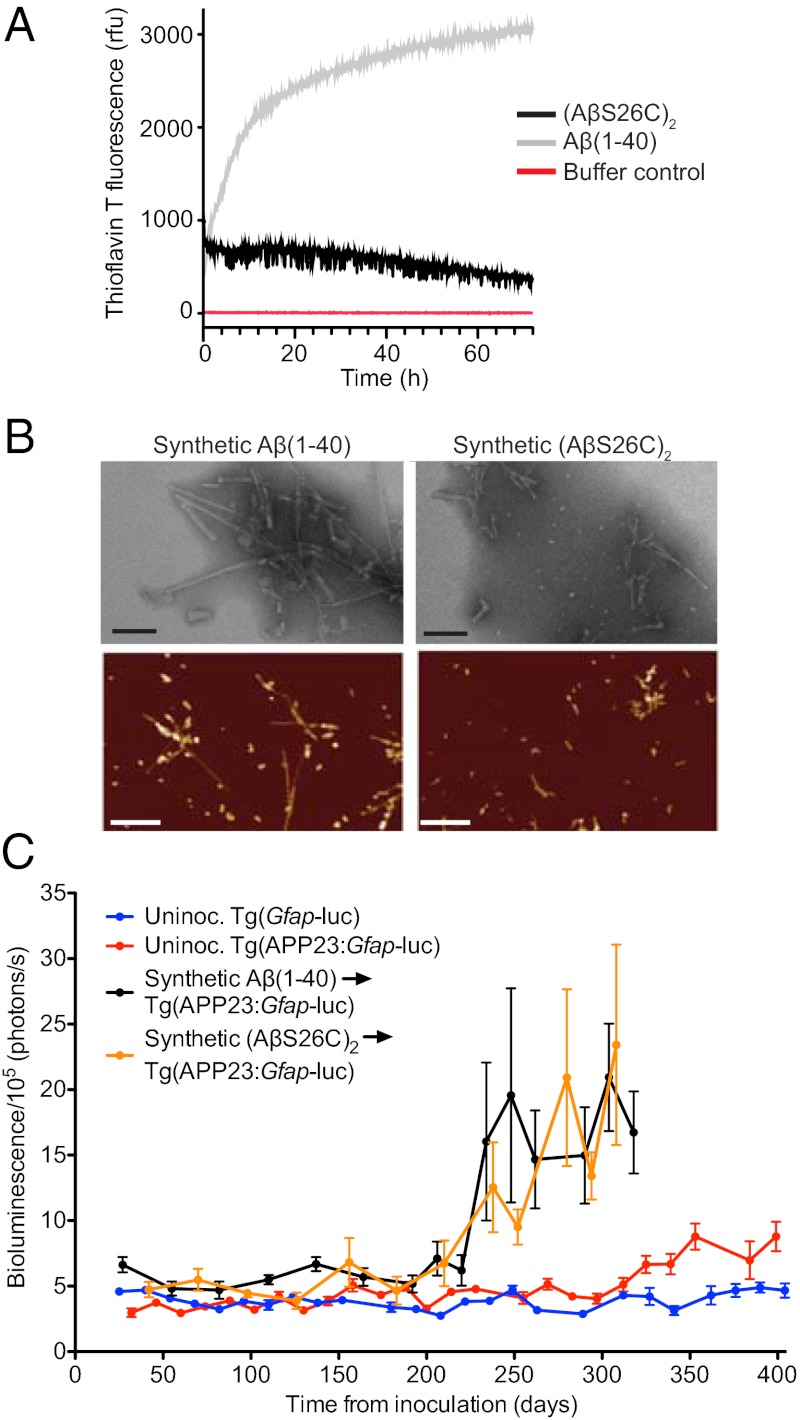
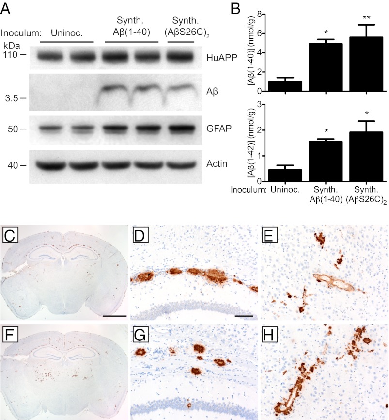
The aggregation and deposition of amyloid-β (Aβ) peptides are believed to be central events in the pathogenesis of Alzheimer's disease (AD). Inoculation of brain homogenates containing Aβ aggregates into susceptible transgenic mice accelerated Aβ deposition, suggesting that Aβ aggregates are capable of self-propagation and hence might be prions. Recently, we demonstrated that Aβ deposition can be monitored in live mice using bioluminescence imaging (BLI). Here, we use BLI to probe the ability of Aβ aggregates to self-propagate following inoculation into bigenic mice. We report compelling evidence that Aβ aggregates are prions by demonstrating widespread cerebral β-amyloidosis induced by inoculation of either purified Aβ aggregates derived from brain or aggregates composed of synthetic Aβ. Although synthetic Aβ aggregates were sufficient to induce Aβ deposition in vivo, they exhibited lower specific biological activity compared with brain-derived Aβ aggregates. Our results create an experimental paradigm that should lead to identification of self-propagating Aβ conformations, which could represent novel targets for interrupting the spread of Aβ deposition in AD patients.


Conflict of interest statement

The authors declare no conflict of interest.

Figures

Fig. 1.
Fig. 1.
Induction of widespread Aβ deposition in Tg(APP23:Gfap-luc) mice inoculated with Aβ amyloid-laden brain homogenate. (A) Mean brain bioluminescence (± SEM) signals obtained from uninoculated Tg(Gfap-luc) mice (blue, n = 16), uninoculated Tg(APP23:Gfap-luc) mice (red, n = 12), Tg(APP23:Gfap-luc) mice inoculated with aged non-Tg brain homogenate (green, n = 6), and Tg(APP23:Gfap-luc) mice inoculated with aged Tg(APP23) (black, n = 6) or Tg(CRND8) (orange, n = 12) brain homogenate. An accelerated increase in the brain BLI signal was observed in mice inoculated with aged Tg(APP23) and Tg(CRND8) brain homogenate compared with mice inoculated with non-Tg brain homogenate and age-matched uninoculated mice. (B) By immunoblotting, increased GFAP and total Aβ levels were apparent in brain homogenates prepared from male Tg(APP23)-inoculated Tg(APP23:Gfap-luc) mice at 385 dpi compared with age-matched mice inoculated with non-Tg brain homogenate. Actin is shown as a control. (C) Widespread induction of Aβ deposition in female Tg(APP23:Gfap-luc) mice inoculated with aged Tg(APP23) brain homogenate at 330 dpi compared with age-matched mice inoculated with brain homogenate from non-Tg mice. Coronal brain sections, cut at the indicated rostral–caudal stereotaxic coordinates, were stained with an anti-Aβ antibody. (Scale bar, 2 mm.)
Fig. 2.
Fig. 2.
Induction of Aβ deposition in Tg(APP23:Gfap-luc) mice inoculated with purified brain-derived Aβ fibrils. (A) Aβ immunoblot (Left) and silver staining (Right) of unpurified brain homogenates (BH) and purified Aβ aggregates (P) from aged Tg(CRND8) and Tg(APP23) mice demonstrated that Aβ peptides are the major protein species after purification. (B) Electron microscopy of purified Aβ fibrils from aged Tg(CRND8) (Left) and Tg(APP23) mice (Right) confirmed the isolation of intact Aβ fibrils. (Scale bars, 100 nm.) (C) Mean brain bioluminescence (± SEM) signals obtained from uninoculated Tg(Gfap-luc) mice (blue, n = 16), uninoculated female Tg(APP23:Gfap-luc) mice (red, n = 6), and female Tg(APP23:Gfap-luc) mice inoculated with Aβ fibrils purified from either aged Tg(APP23) brains (black, n = 11) or aged Tg(CRND8) brains (orange, n = 9). Increased BLI signals were observed in mice inoculated with the purified Aβ fibrils by ∼160 dpi . (D) By immunoblotting, increased GFAP and Aβ protein levels were apparent in brain homogenates prepared from female Tg(APP23:Gfap-luc) mice inoculated with Aβ fibrils purified from aged Tg(APP23) or Tg(CRND8) mice at 300 dpi compared with age-matched, uninoculated mice. Actin levels are shown as a control. (E) By ELISA, Aβ(1–40) (Upper) and Aβ(1–42) (Lower) levels were significantly increased in Tg(APP23:Gfap-luc) mice inoculated with purified Aβ fibrils from Tg(APP23) or Tg(CRND8) mice at 300 dpi compared with age-matched, uninoculated controls (n = 4 each). *P < 0.05, **P < 0.01, ***P < 0.001. Data are mean ± SEM. (FM) Aβ immunostaining of brain sections from female Tg(APP23:Gfap-luc) mice inoculated with purified Tg(APP23) Aβ fibrils (FI) or aged non-Tg brain homogenate (JM) at 300 dpi. Increased Aβ deposition was apparent in whole brain coronal sections (F and J) as well as the hippocampus and overlying cortex (G and K), entorhinal cortex (H and L), and thalamus (I and M) of Aβ-inoculated mice compared with mice inoculated with non-Tg brain homogenate. (Scale bar in F, 2 mm and also applies to J; scale bar in G, 100 μm and also applies to H, I, and KM.)
Fig. 3.
Fig. 3.
Formation and characterization of synthetic Aβ prions. (A) Time course of thioflavin T measurements of Aβ(1–40) (gray line) and (AβS26C)2 (black line) following solubilization, compared with buffer control (red line). (B) Electron microscopy (Upper) (Scale bars, 100 nm.) and atomic force microscopy images (Lower) (Scale bars, 500 nm) of fibrillar aggregates from Aβ(1–40) and (AβS26C)2. (C) Mean brain bioluminescence (± SEM) signals obtained from uninoculated Tg(Gfap-luc) mice (blue, n = 16), uninoculated Tg(APP23:Gfap-luc) mice (red, n = 12), and Tg(APP23:Gfap-luc) mice inoculated with synthetic Aβ aggregates composed of either WT Aβ(1–40) peptide (black, n = 6) or (AβS26C)2 peptide (orange, n = 8). An increase in the BLI signal was observed in mice inoculated with the synthetic Aβ aggregates by ∼230 dpi.
Fig. 4.
Fig. 4.
Induction of cerebral Aβ deposition in Tg(APP23:Gfap-luc) mice inoculated with synthetic Aβ aggregates. (A) Immunoblotting shows that increased GFAP and Aβ protein levels were apparent in brain homogenates prepared from male Tg(APP23:Gfap-luc) mice inoculated with synthetic Aβ aggregates at 330 dpi compared with age-matched, uninoculated Tg(APP23) mice. Actin levels are shown as a control. (B) By ELISA, Aβ(1–40) (Upper) and Aβ(1–42) (Lower) levels were significantly increased in Tg(APP23:Gfap-luc) mice inoculated with synthetic Aβ(1–40) or (AβS26C)2 aggregates at 330 dpi compared with age-matched, uninoculated controls (n = 4 each). *P < 0.05, **P < 0.01. Data are mean ± SEM (CH) Aβ immunostaining of brain sections from female Tg(APP23:Gfap-luc) mice inoculated with synthetic Aβ(1–40) (CE) or (AβS26C)2 (FH) aggregates at 330 dpi. Induced Aβ deposition was apparent in whole brain coronal sections (C and F) as well as the corpus callosum (D and G) and thalamus (E and H) of inoculated mice. (Scale bar in C, 2 mm and also applies to F; scale bar in D, 100 μm and also applies to E, G, and H.)

References

    1. Hardy J, Selkoe DJ. The amyloid hypothesis of Alzheimer’s disease: Progress and problems on the road to therapeutics. Science. 2002;297:353–356. - PubMed
    1. Braak H, Braak E. Neuropathological stageing of Alzheimer-related changes. Acta Neuropathol. 1991;82:239–259. - PubMed
    1. Games D, et al. Alzheimer-type neuropathology in transgenic mice overexpressing V717F β-amyloid precursor protein. Nature. 1995;373:523–527. - PubMed
    1. Hsiao K, et al. Correlative memory deficits, Abeta elevation, and amyloid plaques in transgenic mice. Science. 1996;274:99–102. - PubMed
    1. Sturchler-Pierrat C, et al. Two amyloid precursor protein transgenic mouse models with Alzheimer disease-like pathology. Proc Natl Acad Sci USA. 1997;94:13287–13292. - PMC - PubMed

Publication types

MeSH terms