. 2012 Jul 3;109(27):10891-6.
doi: 10.1073/pnas.1117814109. Epub 2012 Jun 18.

Cell crawling mediates collective cell migration to close undamaged epithelial gaps

Affiliations

Cell crawling mediates collective cell migration to close undamaged epithelial gaps

Ester Anon et al. Proc Natl Acad Sci U S A. .

Abstract

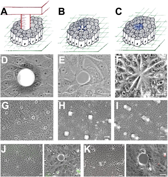
Fundamental biological processes such as morphogenesis and wound healing involve the closure of epithelial gaps. Epithelial gap closure is commonly attributed either to the purse-string contraction of an intercellular actomyosin cable or to active cell migration, but the relative contribution of these two mechanisms remains unknown. Here we present a model experiment to systematically study epithelial closure in the absence of cell injury. We developed a pillar stencil approach to create well-defined gaps in terms of size and shape within an epithelial cell monolayer. Upon pillar removal, cells actively respond to the newly accessible free space by extending lamellipodia and migrating into the gap. The decrease of gap area over time is strikingly linear and shows two different regimes depending on the size of the gap. In large gaps, closure is dominated by lamellipodium-mediated cell migration. By contrast, closure of gaps smaller than 20 μm was affected by cell density and progressed independently of Rac, myosin light chain kinase, and Rho kinase, suggesting a passive physical mechanism. By changing the shape of the gap, we observed that low-curvature areas favored the appearance of lamellipodia, promoting faster closure. Altogether, our results reveal that the closure of epithelial gaps in the absence of cell injury is governed by the collective migration of cells through the activation of lamellipodium protrusion.


Conflict of interest statement

The authors declare no conflict of interest.

Figures

Fig. 1.
Fig. 1.
Experimental design for gap patterning. (AC) Schematic view of the approach used for the experimental model: (A) PDMS pillar surrounded by cells, (B) gap created upon pillar removal, (C) gap closed. (DF) Phase-contrast micrographs of the different stages of the experimental model: (D) PDMS pillar surrounded by cells, (E) gap created upon pillar removal (note that the cells bordering the gap are intact), (F) gap closed by cells. (G) Array of gaps created by using a stencil with numerous PDMS pillars. (H and I) Microfabricated squared (H) and ellipsoidal (I) PDMS pillars with MDCK cells cultured within the pillar stencil. (J) Assessment of cell viability by FITC-dextran uptake. Note that none of the gap lining cells shows positive for FITC-dextran. (K) Assessment of cell viability by propidium iodide internalization. (Scale bars, 10 μm in DF, 20 μm in GK.)
Fig. 2.
Fig. 2.
Dynamics of gap closure. (A) Snapshots of time-lapse video microscopy during gap closure. t = 0 is acquired right after removal of the pillar stencil. (Scale bars, 20 μm.) (B) Evolution of roughness α at the cell–gap interface. Experiments performed with two pillar sizes, 30 and 60 μm in diameter. (C) Gap area decrease with time, for different pillar sizes used for creating the gaps, ranging from 15 to 60 μm in diameter. Green line corresponds to gaps created with pillars of 15 μm in diameter, orange 20, purple 30, black 40, red 50, and blue 60. Data for 80- and 150-μm pillar diameter are shown in Fig. S3. Data are reported as mean and SD of eight gaps analyzed per each size, resulting from four independent experiments. (D) Closure time as a function of the initial gap area of circular gaps. (E) Initial radial velocity depending on the initial gap size. (F) Effect of cell density on closure time, examined for three different gap diameters. Cell density is indicated in the x axis, calculated from Table S2. Each graph shows the experiments from a different pillar diameter. (G) Effect of substrate stiffness on the closure time of two different gap diameters, 20 and 50 μm. The x axis indicates the PDMS ratio used to attain different stiffnesses. No closure stands for gaps that were not closed after 300 min.
Fig. 3.
Fig. 3.
Mechanism of gap closure: effect of inhibitors and actomyosin distribution. (A) Closure times of the different gap sizes in control conditions and subjected to drug treatments of the regulators (MLCK, ROCK, and Rac1 inhibition, for six gap sizes). Data points represent means, and error bars are SEs of seven analyzed gaps. (B) Z-stack projection and xz and yz orthogonal projections (sections from the yellow lines), after 30 min of closure progression Note that there is actin clustering at some cell edges and lamellipodia. (Scale bars, 25 μm in the z-projection, 5 μm in the orthogonal projections.) (C) Actin and phospho-MLC accumulate at the gap margin at t = 0 min but do not progress as a continuous ring as closure proceeds. (Scale bars, 20 μm.) (D) Fold increase in the closure time of Rac-inhibited cells with respect to control conditions for the six different gap sizes.
Fig. 4.
Fig. 4.
Effect of geometry on gap closure. (A) Squared and ellipsoidal PDMS pillars surrounded by cells. (B) Comparison of the closure time of squared and ellipsoidal gaps with respect to circular gaps. (C) Sequence of phase-contrast micrographs showing the progression of the closure of a squared (Top) and ellipsoidal (Bottom) gap. (D and E) Actin (Top) and phospho-MLC (Middle) distribution in squared (D) and ellipsoidal (E) gaps for 2 different time points. (Bottom) Merged images. (F) Epifluorescence micrographs of actin distribution in the closure of ellipsoidal gaps 30 min after pillar removal. (Scale bars: 20 μm.)

References

    1. Martin P, Parkhurst SM. Parallels between tissue repair and embryo morphogenesis. Development. 2004;131:3021–3034. - PubMed
    1. Jacinto A, Martinez-Arias A, Martin P. Mechanisms of epithelial fusion and repair. Nat Cell Biol. 2001;3:E117–E123. - PubMed
    1. Watson AJ, et al. Epithelial barrier function in vivo is sustained despite gaps in epithelial layers. Gastroenterology. 2005;129:902–912. - PubMed
    1. Bement WM, Mandato CA, Kirsch MN. Wound-induced assembly and closure of an actomyosin purse string in Xenopus oocytes. Curr Biol. 1999;9:579–587. - PubMed
    1. Tamada M, Perez TD, Nelson WJ, Sheetz MP. Two distinct modes of myosin assembly and dynamics during epithelial wound closure. J Cell Biol. 2007;176:27–33. - PMC - PubMed

Publication types

LinkOut - more resources