. 2012 Jul 3;109(27):10903-8.
doi: 10.1073/pnas.1118641109. Epub 2012 Jun 18.

Krüppel-like factor 9 is a circadian transcription factor in human epidermis that controls proliferation of keratinocytes

Affiliations

Krüppel-like factor 9 is a circadian transcription factor in human epidermis that controls proliferation of keratinocytes

Florian Spörl et al. Proc Natl Acad Sci U S A. .

Abstract

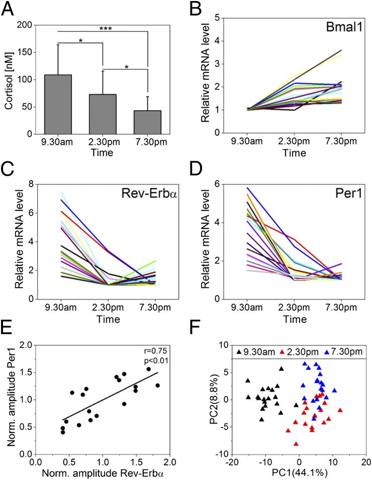
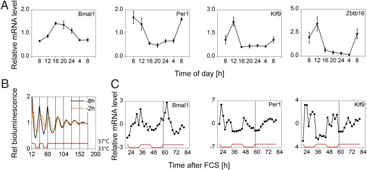
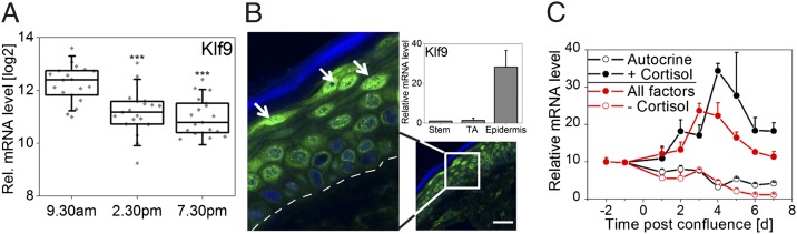
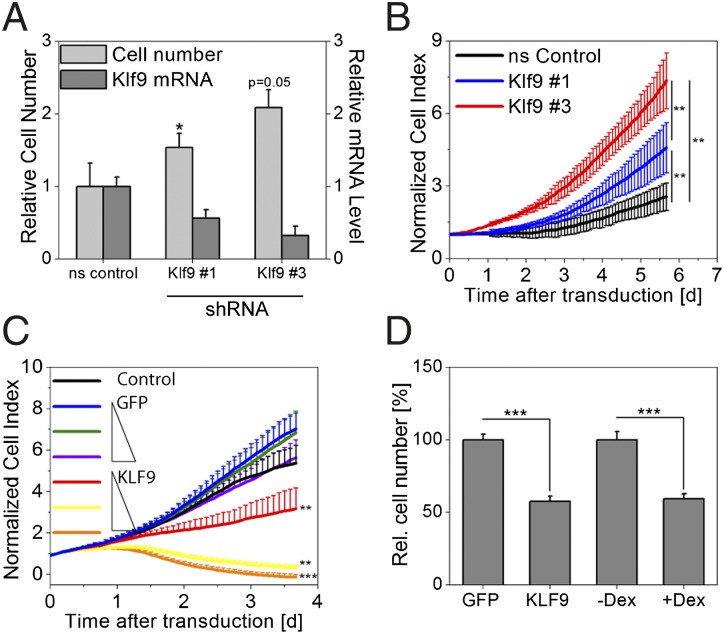
Circadian clocks govern a wide range of cellular and physiological functions in various organisms. Recent evidence suggests distinct functions of local clocks in peripheral mammalian tissues such as immune responses and cell cycle control. However, studying circadian action in peripheral tissues has been limited so far to mouse models, leaving the implication for human systems widely elusive. In particular, circadian rhythms in human skin, which is naturally exposed to strong daytime-dependent changes in the environment, have not been investigated to date on a molecular level. Here, we present a comprehensive analysis of circadian gene expression in human epidermis. Whole-genome microarray analysis of suction-blister epidermis obtained throughout the day revealed a functional circadian clock in epidermal keratinocytes with hundreds of transcripts regulated in a daytime-dependent manner. Among those, we identified a circadian transcription factor, Krüppel-like factor 9 (Klf9), that is substantially up-regulated in a cortisol and differentiation-state-dependent manner. Gain- and loss-of-function experiments showed strong antiproliferative effects of Klf9. Putative Klf9 target genes include proliferation/differentiation markers that also show circadian expression in vivo, suggesting that Klf9 affects keratinocyte proliferation/differentiation by controlling the expression of target genes in a daytime-dependent manner.


Conflict of interest statement

The authors declare no conflict of interest.

Figures

Fig. 1.
Fig. 1.
Circadian gene expression in human epidermis. (A) Cortisol levels in suction-blister liquid obtained at indicated times were determined in 20 individuals. Mean values ± SD are given. *P < 0.05; ***P < 0.001 (Mann–Whitney U test). (BD) Genome-wide microarray analysis was performed using suction-blister epidermis of 19 subjects at indicated times. Bmal1 (B), Rev-Erbα (C), and Per1 (D) expression of each subject are shown relative to minimum. (E) Normalized (relative to mean of all subjects) amplitudes (maximum minus minimum expression) of Per1 and Rev-Erbα were correlated. The Pearson’s correlation coefficient (r = 0.75) and the corresponding P value (P < 0.01) indicate a strong linear correlation. (F) Principal component analysis (PCA) of microarrays was performed for the 250 top-ranking genes according to q value. The relative variance in gene expression (relative to total variance) comprised in PC1 and PC2 is indicated.
Fig. 2.
Fig. 2.
Epidermis clock in vivo and in vitro. (A) Gene expression in punch-biopsy epidermis obtained at indicated times is shown relative to 24-h mean expression (mean of six subjects ± SEM; except Zbtb16: n = 5). Bmal1, Per1, Zbtb16: P < 0.001; Klf9: P = 0.001. (B) Circadian rhythms of Bmal1 reporter activity in primary keratinocytes. Note that instant entrainment is achieved if FCS is administered 8 h before the temperature cycles (black curve). This synchronization paradigm was used for subsequent experiments. (C) Cells were harvested in regular 2-h intervals starting 24 h after FCS for 60 h and mRNA levels of depicted genes were determined. Expression levels are given relative to mean expression. Bmal1, Per1: P < 0.001; Klf9: P = 0.001. (For P values during entrainment and free run, see Table S1C). Statistical analysis for circadian gene expression was performed using CircWave software (43).
Fig. 3.
Fig. 3.
Klf9 is an epidermal differentiation marker. (A) Log2 Klf9 mRNA levels (background-corrected signal intensity) in epidermis of 19 subjects is shown in a box plot. Box, 25–75 percentile; vertical bar, median value; error bars, SDs. Individual signal intensities are shown in gray rectangles. ***P < 0.001 (t test). (B) KLF9 protein levels in human skin section determined by immunofluorescence staining. KLF9 immunoreactivity is shown in green and nuclei are stained with DAPI (blue). The stratum corneum layer (outermost epidermal layer) is indicated in blue, and the basal membrane is indicated as a white dashed line. Arrowheads show strong staining in differentiated cells in upper epidermal layers (stratum granulosum). (Scale bar: 50 μm.) (Inset) Klf9 mRNA levels in validated cell fractions (23) of stem cells, transient amplifying keratinocytes (TA Cell), and fully differentiated epidermis (Epidermis) and given relative to the stem cell fraction (four subjects; mean ± SD). (C) Keratinocytes were differentiated by cultivating cells into postconfluence using different culture conditions: full growth medium ± cortisol (All factors and -Cortisol, respectively) or autocrine growth conditions ± cortisol (Autocrine and +Cortisol, respectively). Klf9 mRNA levels of two experiments (mean ± maximum or minimum) are shown relative to minimum expression.
Fig. 4.
Fig. 4.
Klf9 attenuates keratinocyte proliferation. (A) Klf9 transcript was down-regulated in primary keratinocytes using stably integrated shRNAmir constructs (#1 and #3), and cell numbers were assessed 5 d after transduction. Klf9 mRNA levels and cell numbers are given relative to cells transduced with a nonsilencing (ns control) vector (mean ± SD of three experiments; *P < 0.05, t test). (B) Cells were treated similar to A, and cell growth was assessed using real-time impedance measurements (mean value of six wells ± SD; representative plot of three experiments is shown). (C) Primary keratinocytes were transduced with increasing (1×, 2×, and 4×) viral titers carrying GFP or KLF9 expression constructs or left untreated (Control). Cell growth was subsequently analyzed in real-time impedance measurements (mean value of six wells ± SD; representative plot of three experiments is shown). **P < 0.01; ***P < 0.001 (Mann–Whitney U test). (D) Cells were treated similar to C, and a different set of cells was treated with dex. Cell number was analyzed by cell counting 4 d after transduction/treatment [1× virus; mean ± SEM of 9 wells (virus) or 24 wells (dex treatment) of two pooled independent experiments; ***P < 0.01, t test).

References

    1. Reppert SM, Weaver DR. Coordination of circadian timing in mammals. Nature. 2002;418:935–941. - PubMed
    1. Dibner C, Schibler U, Albrecht U. The mammalian circadian timing system: Organization and coordination of central and peripheral clocks. Annu Rev Physiol. 2010;72:517–549. - PubMed
    1. Fonjallaz P, Ossipow V, Wanner G, Schibler U. The two PAR leucine zipper proteins, TEF and DBP, display similar circadian and tissue-specific expression, but have different target promoter preferences. EMBO J. 1996;15:351–362. - PMC - PubMed
    1. Nakahata Y, Sahar S, Astarita G, Kaluzova M, Sassone-Corsi P. Circadian control of the NAD+ salvage pathway by CLOCK-SIRT1. Science. 2009;324:654–657. - PMC - PubMed
    1. Schibler U. Circadian time keeping: The daily ups and downs of genes, cells, and organisms. Prog Brain Res. 2006;153:271–282. - PubMed

Publication types

MeSH terms

Associated data