. 2012 Jul 10;109(28):E1938-46.
doi: 10.1073/pnas.1206999109. Epub 2012 Jun 18.

Cofactor molecules maintain infectious conformation and restrict strain properties in purified prions

Affiliations

Cofactor molecules maintain infectious conformation and restrict strain properties in purified prions

Nathan R Deleault et al. Proc Natl Acad Sci U S A. .

Abstract

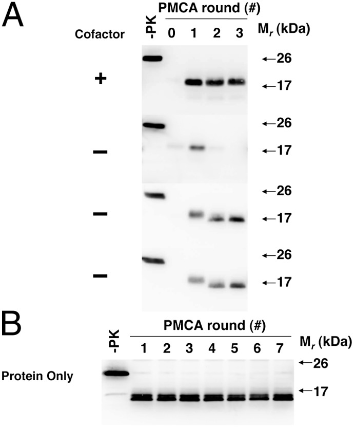
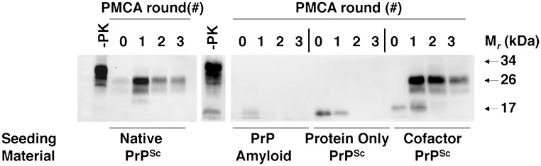
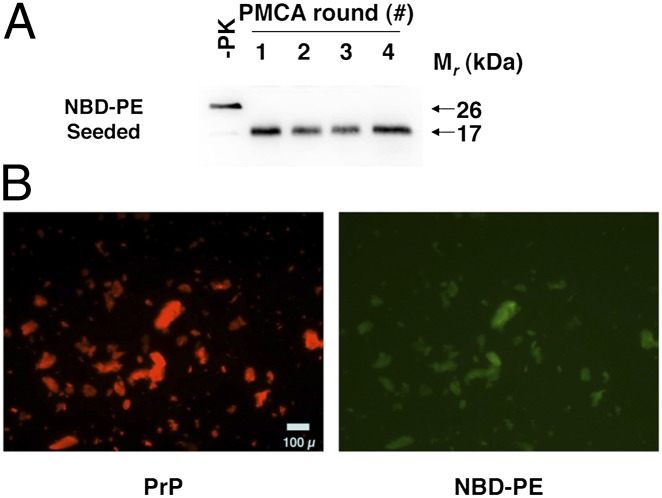
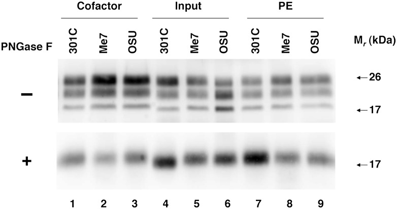
Prions containing misfolded prion protein (PrP(Sc)) can be formed with cofactor molecules using the technique of serial protein misfolding cyclic amplification. However, it remains unknown whether cofactors materially participate in maintaining prion conformation and infectious properties. Here we show that withdrawal of cofactor molecules during serial propagation of purified recombinant prions caused adaptation of PrP(Sc) structure accompanied by a reduction in specific infectivity of >10(5)-fold, to undetectable levels, despite the ability of adapted "protein-only" PrP(Sc) molecules to self-propagate in vitro. We also report that changing only the cofactor component of a minimal reaction substrate mixture during serial propagation induced major changes in the strain properties of an infectious recombinant prion. Moreover, propagation with only one functional cofactor (phosphatidylethanolamine) induced the conversion of three distinct strains into a single strain with unique infectious properties and PrP(Sc) structure. Taken together, these results indicate that cofactor molecules can regulate the defining features of mammalian prions: PrP(Sc) conformation, infectivity, and strain properties. These findings suggest that cofactor molecules likely are integral components of infectious prions.


Conflict of interest statement

Conflict of interest statement: S.S. and N.D. are inventors on a patent held by Dartmouth College on the use of phosphatidylethanolamine as a prion cofactor.

Figures

Fig. 1.
Fig. 1.
Adaptation of autocatalytic PrPSc molecules. Western blots of reconstituted sPMCA reactions. −PK, samples not subjected to proteinase K digestion; all other samples were proteolyzed. (A) All reactions initially were seeded with OSU cofactor PrPSc molecules and subsequently were propagated in substrate mixtures with or without cofactor, as indicated. (B) Ongoing propagation of OSU protein-only PrPSc molecules in substrate mixture lacking cofactor.
Fig. 2.
Fig. 2.
Seeding of brain homogenate sPMCA reactions. Western blot of three-round sPMCA reactions using normal mouse brain homogenate substrate seeded with various samples as indicated. −PK, samples not subjected to proteinase K digestion; all other samples were proteolyzed. Native PrPSc was isolated as PrP27-30 molecules from the brains of Me7-infected mice as previously described (35), and PrP amyloid was generated as previously described (36).
Fig. 3.
Fig. 3.
NBD-PE PrPSc colocalization assay. (A) Western blot of a four-round sPMCA reaction using recPrP and NBD-PE as substrate. −PK, samples not subjected to proteinase K digestion; all other samples were proteolyzed. (B) Dual-channel fluorescence micrographs showing representative images of the final product of the sPMCA reaction shown in A, after purification with detergent washes as described in Experimental Procedures. PrPSc aggregates immunostained with anti-PrP mAb D13 and Alexa Fluor 568-labeled secondary antibody are shown in red (PrP), and colocalized NBD-PE molecules are shown in green (NBD-PE).
Fig. 4.
Fig. 4.
Regional neuropathology of infected mice. (AD) Profiles of vacuolation scores of animals inoculated with samples containing (A) input prions, (B) cofactor PrPSc molecules, (C) PE PrPSc prions, or (D) serial-passage PE PrPSc prions. (EH) Profiles of PrP deposition scores of animals inoculated with samples containing (E) input prions, (F) cofactor PrPSc molecules, (G) PE PrPSc prions, or (H) serial-passage PE PrPSc prions. Prion strains: OSU, red squares; Me7, blue circles; 301C, green triangles. Brain regions: I–II, cerebral cortical layers 1 and 2; III–IV, cortical layers 3 and 4; V–VI, cortical layers 5 and 6; BS, brainstem; Cb, cerebellum; CC, cerebral cortex (all layers); H, hippocampus; HT, hypothalamus; Mid, midbrain; T, thalamus. Mean values ± SEM are shown; n = 5.
Fig. 5.
Fig. 5.
Glycoform distribution and electrophoretic mobility of PrPSc molecules in the brains of infected mice. (Upper) Western blots of brain homogenate samples prepared from animals inoculated with samples containing input prions, cofactor PrPSc, and PE PrPSc molecules derived from different prion strains, as indicated. All samples were subjected to limited proteolysis. (Lower) Samples also were deglycosylated by treatment with PNGase F, as indicated (+), before SDS/PAGE.
Fig. 6.
Fig. 6.
Analysis of PrPSc conformational stability. Urea denaturation assay showing PrPSc levels in samples of brain homogenates prepared from animals inoculated with samples derived from different prion strains. Inocula were (A) input prions; (B) cofactor PrPSc; (C) PE PrPSc; (D) serial-passage PE PrPSc. OSU, red squares; Me7, blue circles; 301C, green triangles. Mean values ± SEM of three replicates are shown for each point.
Fig. P1.
Fig. P1.
Cofactor variation hypothesis: Multiple cofactors generate strain diversity, represented by the different geometric shapes of each PrPSc molecule.

References

    1. Prusiner SB. Novel proteinaceous infectious particles cause scrapie. Science. 1982;216:136–144. - PubMed
    1. Caughey BW, et al. Secondary structure analysis of the scrapie-associated protein PrP 27-30 in water by infrared spectroscopy. Biochemistry. 1991;30:7672–7680. - PubMed
    1. Pan KM, et al. Conversion of alpha-helices into beta-sheets features in the formation of the scrapie prion proteins. Proc Natl Acad Sci USA. 1993;90:10962–10966. - PMC - PubMed
    1. Kocisko DA, et al. Cell-free formation of protease-resistant prion protein. Nature. 1994;370:471–474. - PubMed
    1. Deleault NR, Harris BT, Rees JR, Supattapone S. Formation of native prions from minimal components in vitro. Proc Natl Acad Sci USA. 2007;104:9741–9746. - PMC - PubMed

Publication types