. 2012 Jul 15;189(2):516-20.
doi: 10.4049/jimmunol.1200064. Epub 2012 Jun 18.

Cutting edge: impaired MHC class I expression in mice deficient for Nlrc5/class I transactivator

Affiliations

Cutting edge: impaired MHC class I expression in mice deficient for Nlrc5/class I transactivator

Amlan Biswas et al. J Immunol. .

Abstract

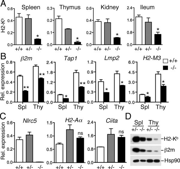
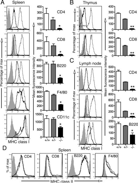
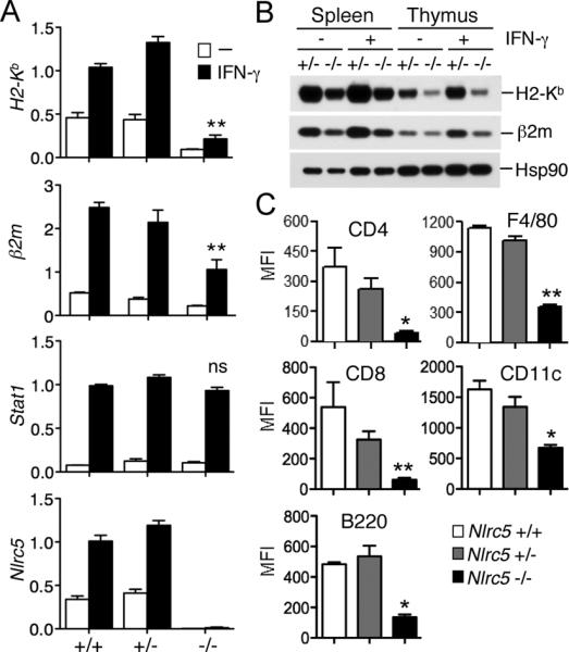
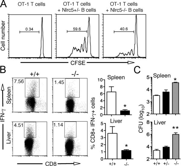
MHC class I and class II are crucial for the adaptive immune system. Although regulation of MHC class II expression by CIITA has long been recognized, the mechanism of MHC class I transactivation has been largely unknown until the recent discovery of NLRC5/class I transactivator. In this study, we show using Nlrc5-deficient mice that NLRC5 is required for both constitutive and inducible MHC class I expression. Loss of Nlrc5 resulted in severe reduction in the expression of MHC class I and related genes such as β(2)-microglobulin, Tap1, or Lmp2, but did not affect MHC class II levels. IFN-γ stimulation could not overcome the impaired MHC class I expression in Nlrc5-deficient cells. Upon infection with Listeria monocyogenes, Nlrc5-deficient mice displayed impaired CD8(+) T cell activation, accompanied with increased bacterial loads. These findings illustrate critical roles of NLRC5/class I transactivator in MHC class I gene regulation and host defense by CD8(+) T cell responses.


Figures

FIGURE 1
FIGURE 1
Nlrc5 is required for the constitutive expression of MHC class I in various organs. (A) Expression of MHC class I (H2-Kb) in the indicated organs of wild-type mice (+/+, n=3), or mice heterozygous (+/−, n=4) or homozygous (−/−, n=3) for the targeted disruption of the Nlrc5 gene, as determined by qPCR analysis using gene-specific primers. Error bars represent ± SEM. *p < 0.05. (B) Expression of β2m, Tap1, Lmp2 and H2-M3 in the spleen and thymus of Nlrc5+/+ (n = 3) and Nlrc5−/− mice (n = 3) were analyzed by qPCR using the indicated gene-specific primers. Error bars represent ± SEM. *p < 0.05; **p < 0.01; ns, not significant. (C) Analysis of Nlrc5, MHC class II (H2-Aa), and CIITA gene expression in splenocytes, as determined by qPCR. n = 3. Error bars represent ± SEM. ns: not significant. (D) Western blot analysis of MHC class I (H2-Kb) and β2m protein expression in the spleen and thymus from mice with the indicated genotype. The expression of Hsp90 is shown as a loading control.
FIGURE 2
FIGURE 2
Reduced MHC class I expression in various cell types of Nlrc5-deficient mice. Single cell suspension of spleen (A, left panel & D), thymus (B, left panel) and lymph node (C, left panel) from Nlrc5 +/− (grey line) and Nlrc5−/− (black line) mice were analyzed by flow cytometry for the expression of MHC class I (H2-K/D/L, in A, B & C, left panel) or MHC class II (D) molecules (IAb/IAd), gated on CD8+, CD4+, B220+, F4/80+ or CD11c+ cells. Shaded region represent the isotype control. Data are representative of two independent experiments. Bar graphs (A, B & C, right panel) present the mean and SEM of the corresponding MFI (n = 3 for each genotype). *p < 0.05; **p < 0.01.
FIGURE 3
FIGURE 3
IFN-γ stimulation does not rescue the reduced MHC class I expression observed in Nlrc5-deficient mice. (A) Splenocytes from Nlrc5+/+, Nlrc5+/− and Nlrc5−/−mice (n = 3 for each genotype) were stimulated for 18 hrs with IFN-γ (100 U/ml) and transcript levels were analyzed by qPCR using the indicated gene-specific primers. Error bars represent ± SEM. **p < 0.01; ns, not significant. (B) MHC class I (H2-Kb) and β2m protein expression in splenocytes and thymocytes from mice with the indicated genotype. Cell extracts were prepared 18 hrs post stimulation with IFN-γ and analyzed by Western blotting. Hsp90 levels are depicted as a loading control. (C) Splenocytes from Nlrc5+/+, Nlrc5+/− and Nlrc5−/− mice (n = 3 for each genotype) were cultured overnight in the presence of IFN-γ and were analyzed by flow cytometry for the expression of MHC class I molecules (H2-K/D/L), gated on CD8+, CD4+, B220+, F4/80+, and CD11c+ cells. Bar graphs represent the mean and SEM of the corresponding MFI.
FIGURE 4
FIGURE 4
Nlrc5 is required for MHC class I mediated immune responses. (A) Nlrc5-deficient B cells display impaired antigen-specific stimulation of CD8+ T cells. CFSE-labeled OT-1 (CD45.1) CD8+ T cells were co-cultured with SIINFEKL (1μM) pulsed B220+ splenocytes from Nlrc5+/− or Nlrc5−/− mice at a ratio of 1:25 (B:T) for 72 hrs. Proliferation of CD45.1 gated cells was determined by flow cytometric analysis of CFSE dilution. Data are representative of two independent experiments (n=3). (B) Nlrc5+/+, Nlrc5+/− or Nlrc5−/− mice were infected with L. monocytogenes (LM) (1 × 10⁴ CFU). Splenocytes and hepatic leucocytes isolated from Nlrc5+/+ and Nlrc5−/− mice at day 6 after infection were cultured with heat-killed LM for 16 hours and analyzed by flow cytometry for the expression of IFN-γ gated on CD8+ cells. Numbers indicate percentage of cells. Data are representative of two independent experiments (n=3). Bar graphs (B, right panel) present the mean and SEM of the percentage of CD8+ IFN-γ+ cells (n = 3 for each genotype). *p < 0.05. (C) Bacterial load in the spleen and liver of LM infected Nlrc5+/+, Nlrc5+/− or Nlrc5−/− mice were determined. Data are representative of two independent experiments *p < 0.05; **p < 0.01.

References

    1. Germain RN, Margulies DH. The biochemistry and cell biology of antigen processing and presentation. Annu. Rev. Immunol. 1993;11:403–450. - PubMed
    1. Steimle V, Otten LA, Zufferey M, Mach B. Complementation cloning of an MHC class II transactivator mutated in hereditary MHC class II deficiency (or bare lymphocyte syndrome) Cell. 1993;75:135–146. - PubMed
    1. Beresford GW, Boss JM. CIITA coordinates multiple histone acetylation modifications at the HLA-DRA promoter. Nat. Immunol. 2001;2:652–657. - PubMed
    1. Masternak K, Muhlethaler-Mottet A, Villard J, Zufferey M, Steimle V, Reith W. CIITA is a transcriptional coactivator that is recruited to MHC class II promoters by multiple synergistic interactions with an enhanceosome complex. Genes Dev. 2000;14:1156–1166. - PMC - PubMed
    1. Reith W, Mach B. The bare lymphocyte syndrome and the regulation of MHC expression. Annu. Rev. Immunol. 2001;19:331–373. - PubMed

Publication types