. 2012 Sep;53(9):1840-50.
doi: 10.1194/jlr.M024471. Epub 2012 Jun 18.

Quercetin enhances ABCA1 expression and cholesterol efflux through a p38-dependent pathway in macrophages

Affiliations

Quercetin enhances ABCA1 expression and cholesterol efflux through a p38-dependent pathway in macrophages

Yu-Cheng Chang et al. J Lipid Res. 2012 Sep.

Abstract

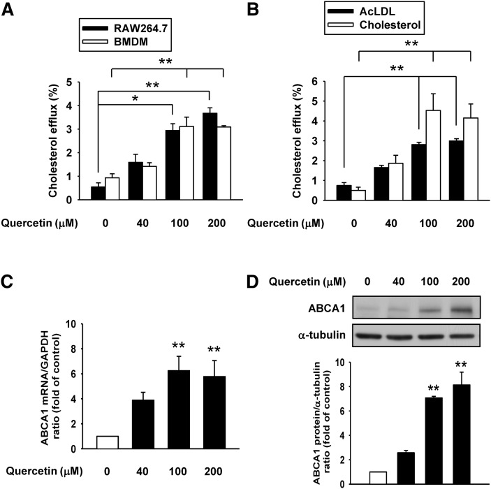
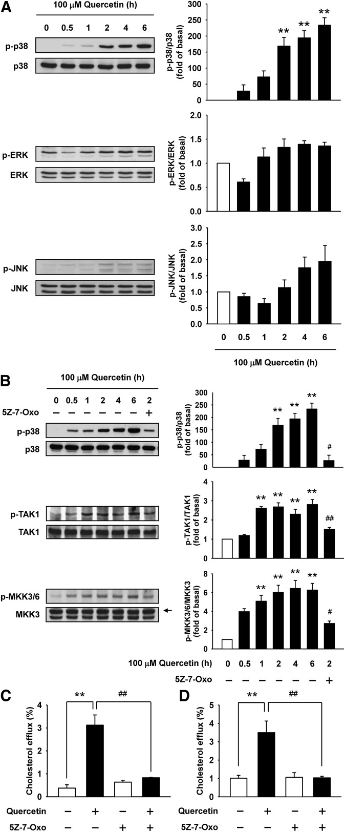
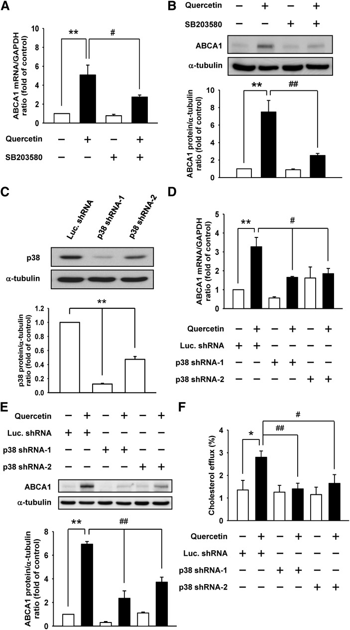
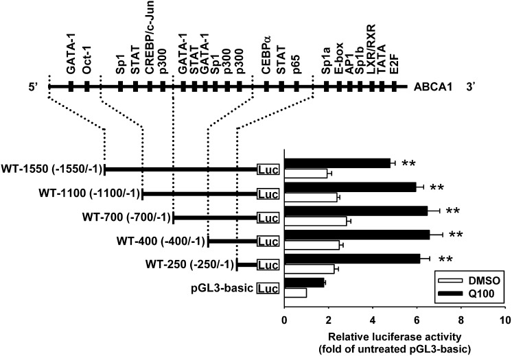
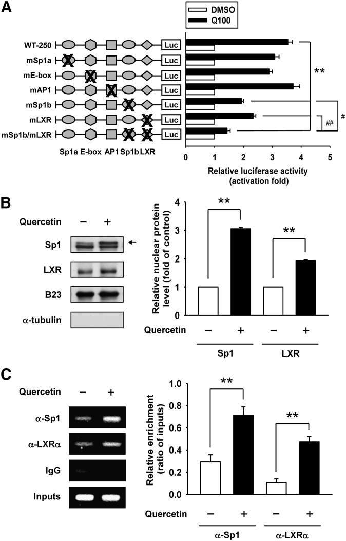
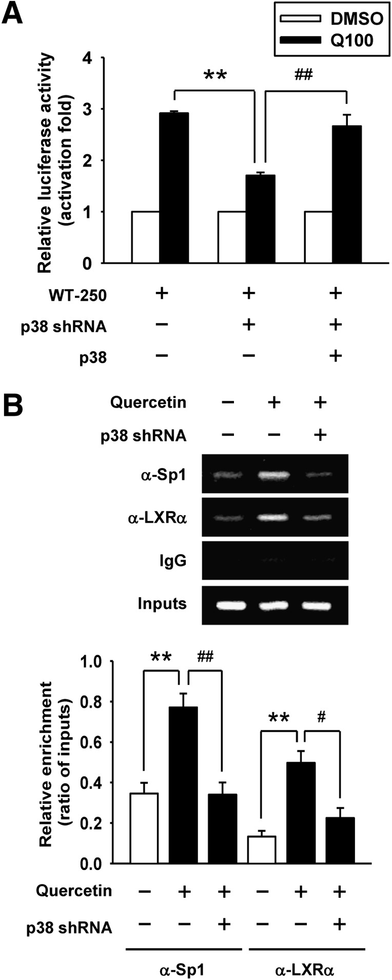
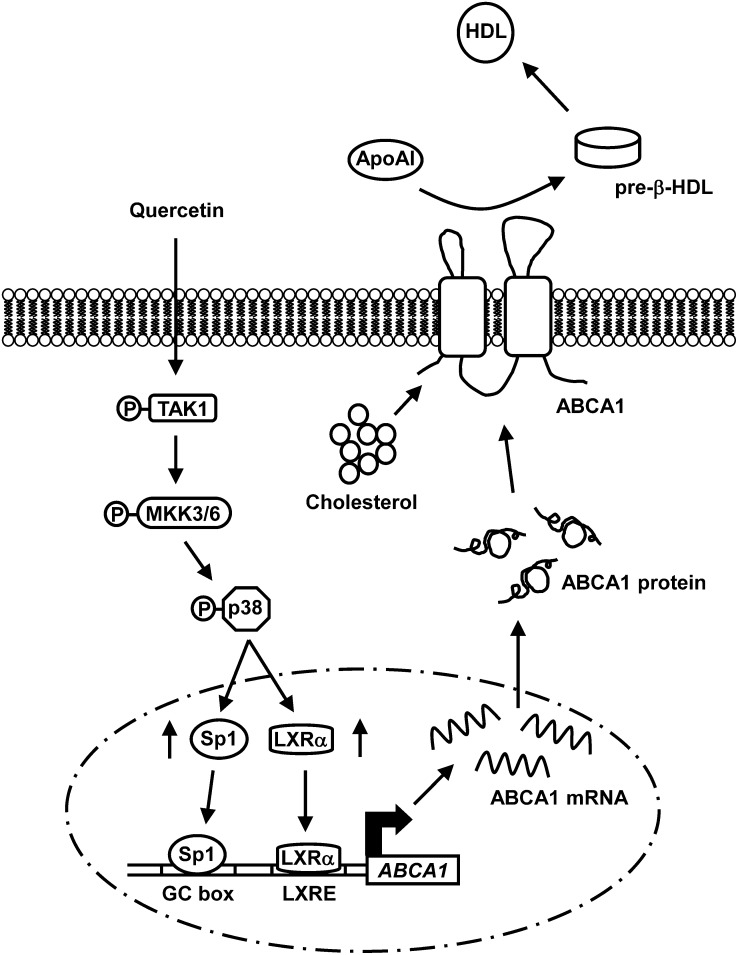
ATP-binding cassette transporter A1 (ABCA1) plays a crucial role in exporting cholesterol from macrophages, a function relevant to its involvement in the prevention of atherosclerosis. Quercetin, one of flavonoids, has been described to reduce atherosclerotic lesion formation. This study is aimed to investigate the effect of quercetin on regulation of ABCA1 expression and to explore its underlying mechanisms in macrophages. The results show that quercetin markedly enhanced cholesterol efflux from macrophages in a concentration-dependent manner, which was associated with an increase in ABCA1 mRNA and protein expression. Remarkably, quercetin is able to stimulate the phosphorylation of p38 by up to 234-fold at 6 h via an activation of the transforming growth factor β-activated kinase 1 (TAK1) and mitogen-activated kinase kinase 3/6 (MKK3/6). Inhibition of p38 with a pharmacological inhibitor or small hairpin RNA (shRNA) suppressed the stimulatory effects of quercetin on ABCA1 expression and cholesterol efflux. Moreover, knockdown of p38 reduced quercetin-enhanced ABCA1 promoter activity and the binding of specificity protein 1 (Sp1) and liver X receptor α (LXRα) to the ABCA1 promoter using chromatin immunoprecipitation assays. These findings provide evidence that p38 signaling is essential for the regulation of quercetin-induced ABCA1 expression and cholesterol efflux in macrophages.


Figures

Fig. 1.
Fig. 1.
Quercetin enhances cholesterol efflux and expression of ABCA1 in macrophages. A: [3H]cholesterol-labeled RAW264.7 macrophages or BMDM were treated with quercetin as indicated concentrations for 24 h. ApoAI-dependent cholesterol efflux was measured by incubating [3H]cholesterol-labeled macrophages with or without 10 μg/ml apoAI for 24 h. Cholesterol efflux was expressed as the percentage of radioactivity in the medium relative to the total radioactivity (medium and cells). B: RAW264.7 macrophages were loaded with 10 μg/ml of acetylated low-density lipoprotein (AcLDL) or 30 μg/ml cholesterol in the presence of [3H]cholesterol for 24 h. Cholesterol efflux was measured as described in Materials and Methods. C: After cells were treated with quercetin for 18 h, mRNA levels of ABCA1 were detected by quantitative real-time PCR and normalized to GAPDH. D: Cells were treated with quercetin for 24 h, and then total cell extracts were harvested. ABCA1 protein expressions were measured by Western blot analysis and α-tubulin was utilized as a loading control. The normalized level of mRNA or protein from cells without quercetin treatment was set as 1. Results are expressed as mean ± SEM (n = 3–5). *P < 0.05, **P < 0.01 versus control group.
Fig. 2.
Fig. 2.
Quercetin activates p38 via the TAK1-MKK3/6 signaling cascade. A: The proteins from total cell lysates were separated by SDS-PAGE and immunoblotted with anti-phospho-MAPK antibodies. Immunoblots were reprobed with total MAPK antibodies for internal normalization. B: RAW264.7 macrophages were pretreated with 5 μM 5Z-7-Oxo for 1 h, and then treated with 100 μM quercetin as indicated time to determine whether the p38 activation was via phosphorylation of TAK1 and MKK3/6. An arrow indicates MKK3 band. Histograms show the relative intensity of normalized phospho-MAPK, phospho-TAK1, and phospho-MKK3/6 over the 0 h group. Data represent mean ± SEM (n = 3–5). **P < 0.01 versus 0 h group, #P < 0.05 versus 2 h group. C, D: Analyses were performed to determine the effect of 5Z-7-Oxo on cholesterol efflux in RAW264.7 macrophages (C) or BMDM (D) as described in Materials and Methods. Bars are mean ± SEM (n = 3–4). **P < 0.01, ##P < 0.01.
Fig. 3.
Fig. 3.
p38 is involved in the quercetin-induced ABCA1 expression and cholesterol efflux. RAW264.7 macrophages were pretreated with 20 μM SB203580 (p38 inhibitor), and then treated with 100 μM quercetin. A: The ABCA1 mRNA levels were measured by quantitative real-time PCR and normalized to GAPDH. B: The ABCA1 protein levels were detected by Western blot analysis and α-tubulin was utilized as a loading control. The normalized level of mRNA or protein from cells without quercetin treatment was set as 1. RAW264.7 macrophages were infected with lentivirus expressing p38 shRNA-1 or p38 shRNA-2 to confirm the effects of p38 on quercetin-induced ABCA1 expression and cholesterol efflux. Luciferase shRNA (Luc. shRNA) was used as a control shRNA. C: The knockdown efficiency of p38 was checked by Western blot analysis. D, E: The effect of p38 knockdown on quercetin-induced ABCA1 mRNA (D) and protein (E) expression was examined by quantitative real-time PCR and Western blot analysis, respectively. The normalized level of mRNA or protein from parental cells without quercetin treatment was given as 1. F: Cholesterol efflux from p38 knockdown cells to media was measured in the presence of 10 μg/ml apoAI. Cholesterol efflux was expressed as the percentage of radioactivity in the medium relative to the total radioactivity (medium and cells). Values are mean ± SEM (n = 3–6). *P < 0.05, **P < 0.01, #P < 0.05, ##P < 0.01.
Fig. 4.
Fig. 4.
Characterization of quercetin-responsive domains in the ABCA1 promoter. Schematic diagram of putative binding sites for transcription factors and serial deletion constructs of ABCA1 promoter are shown. RAW264.7 macrophages were transiently cotransfected with 1 μg of indicated constructs and 0.5 μg of pCMV β-galactosidase expression plasmid, and then treated with 100 μM quercetin (Q100) for 24 h. Luciferase activity was normalized with β-galactosidase activity. Results are shown as fold changes in luciferase activity relative to the untreated pGL3-basic vector group. The level of luciferase activity without quercetin treatment in pGL3-basic vector group was given the value of 1. Bars are mean ± SEM (n = 3). **P < 0.01 versus quercetin-treated pGL3-basic vector group.
Fig. 5.
Fig. 5.
Sp1 and LXR binding sites are quercetin-responsive elements within the ABCA1 promoter. A: Six mutation constructs of ABCA1 promoter in the potential Sp1a, E-box, AP1, Sp1b, and LXR recognition sites were generated using site-directed mutatgenesis. RAW264.7 macrophages were transiently cotransfected with 1 μg of indicated mutant constructs and 0.5 μg of pCMV β-galactosidase expression plasmid, and then treated with 100 μM quercetin (Q100) for 24 h. Luciferase activity was normalized with β-galactosidase activity and expressed as fold changes in luciferase activity compared with those of the control. The level of luciferase activity without quercetin treatment was given the value of 1. WT-250, −250/−1 region of ABCA1 promoter. mSp1b/mLXR, double mutations at Sp1b and LXR sites. B: Cells were treated with quercetin for 3 h, and nuclear extracts were harvested. The nuclear expression levels of Sp1 and LXR were analyzed by Western blot analysis. The arrow indicates Sp1 band. B23 was used as nuclear loading control, and α-tubulin was used to exclude cytosolic contamination. The level of Sp1 or LXR without quercetin treatment was given the value of 1. C: ChIP assays were performed to observe the binding of Sp1 and LXRα to the ABCA1 promoter in quercetin-treated cells as described in Materials and Methods. Nonimmune IgG was used as the negative control. The results of the ChIP assays were evaluated by PCR and gel electrophoresis. Values are quantified by densitometer and expressed as ratio relative to inputs. Results are mean ± SEM (n = 3–4). **P < 0.01, #P < 0.05, ##P < 0.01.
Fig. 6.
Fig. 6.
Effect of p38 knockdown by shRNA on the attenuation of Sp1 and LXRα binding to the ABCA1 promoter. A: Parental cells or p38 knockdown cells were transiently cotransfected with 0.5 μg of WT-250 construct, 0.5 μg of pRcCMV vector, and 0.5 μg of pCMV β-galactosidase plasmid. For p38 overexpression, p38 knockdown cells were transiently cotransfected with 0.5 μg of WT-250 construct, 0.5 μg of pRcCMV-p38 plasmid, and 0.5 μg of pCMV β-galactosidase plasmid. After transfection, cells were treated with 100 μM quercetin (Q100) for 24 h. Luciferase activity was normalized with β-galactosidase activity and expressed as fold changes in luciferase activity compared with respective control groups. The level of luciferase activity without quercetin treatment was given the value of 1. WT-250, −250/−1 region of ABCA1 promoter. B: ChIP assays were performed to observe the binding of Sp1 and LXRα to the ABCA1 promoter in p38 knockdown cells as described in Materials and Methods. Nonimmune IgG was used as the negative control. The results of ChIP assays were evaluated by PCR and gel electrophoresis. Values are quantified by densitometer and expressed as ratio relative to inputs. Bars are mean ± SEM (n = 3). **P < 0.01, #P < 0.05, ##P < 0.01.
Fig. 7.
Fig. 7.
A model describing the mechanisms of quercetin-induced cholesterol efflux in macrophages. Quercetin elicits the TAK1-MKK3/6 signaling cascade to activate p38. Activated p38 subsequently increases the binding of Sp1 and LXRα to the ABCA1 promoter, which in turn enhances the expression of ABCA1 as well as cholesterol efflux from macrophages.

Similar articles

Cited by

References

    1. Peluso M. R. 2006. Flavonoids attenuate cardiovascular disease, inhibit phosphodiesterase, and modulate lipid homeostasis in adipose tissue and liver. Exp. Biol. Med. (Maywood). 231: 1287–1299. - PubMed
    1. Boots A. W., Haenen G. R., Bast A. 2008. Health effects of quercetin: from antioxidant to nutraceutical. Eur. J. Pharmacol. 585: 325–337. - PubMed
    1. Bischoff S. C. 2008. Quercetin: potentials in the prevention and therapy of disease. Curr. Opin. Clin. Nutr. Metab. Care. 11: 733–740. - PubMed
    1. Kleemann R., Verschuren L., Morrison M., Zadelaar S., van Erk M. J., Wielinga P. Y., Kooistra T. 2011. Anti-inflammatory, anti-proliferative and anti-atherosclerotic effects of quercetin in human in vitro and in vivo models. Atherosclerosis. 218: 44–52. - PubMed
    1. Rivera L., Moron R., Sanchez M., Zarzuelo A., Galisteo M. 2008. Quercetin ameliorates metabolic syndrome and improves the inflammatory status in obese Zucker rats. Obesity (Silver Spring). 16: 2081–2087. - PubMed

Publication types

MeSH terms