

A Neural Theory of Speech Acquisition and Production
- PMID: 22711978
- PMCID: PMC3375605
- DOI: 10.1016/j.jneuroling.2009.08.006
A Neural Theory of Speech Acquisition and Production
Abstract
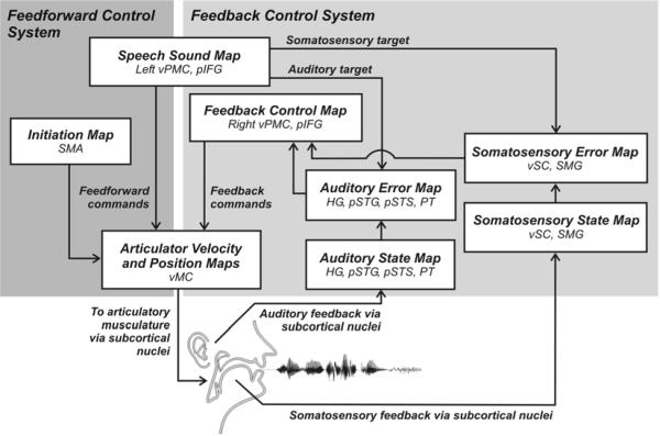
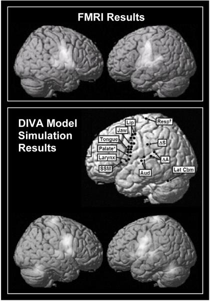
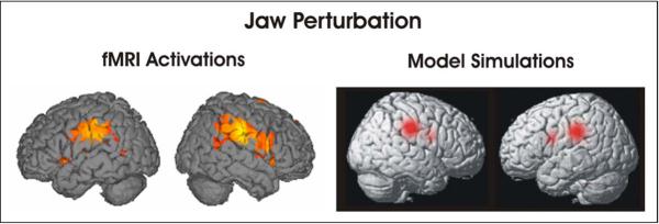
This article describes a computational model, called DIVA, that provides a quantitative framework for understanding the roles of various brain regions involved in speech acquisition and production. An overview of the DIVA model is first provided, along with descriptions of the computations performed in the different brain regions represented in the model. Particular focus is given to the model's speech sound map, which provides a link between the sensory representation of a speech sound and the motor program for that sound. Neurons in this map share with "mirror neurons" described in monkey ventral premotor cortex the key property of being active during both production and perception of specific motor actions. As the DIVA model is defined both computationally and anatomically, it is ideal for generating precise predictions concerning speech-related brain activation patterns observed during functional imaging experiments. The DIVA model thus provides a well-defined framework for guiding the interpretation of experimental results related to the putative human speech mirror system.
Figures

Similar articles
-
The DIVA model: A neural theory of speech acquisition and production.Lang Cogn Process. 2011 Jan 1;26(7):952-981. doi: 10.1080/01690960903498424. Lang Cogn Process. 2011. PMID: 23667281 Free PMC article.
-
Modelling speech motor programming and apraxia of speech in the DIVA/GODIVA neurocomputational framework.Aphasiology. 2021;35(4):424-441. doi: 10.1080/02687038.2020.1765307. Epub 2020 May 18. Aphasiology. 2021. PMID: 34108793 Free PMC article.
-
On the context-dependent nature of the contribution of the ventral premotor cortex to speech perception.Neuroimage. 2011 Aug 15;57(4):1561-71. doi: 10.1016/j.neuroimage.2011.05.067. Epub 2011 Jun 1. Neuroimage. 2011. PMID: 21664275 Free PMC article.
-
Neurocomputational modeling of speech motor development.J Child Lang. 2023 Nov;50(6):1318-1335. doi: 10.1017/S0305000923000260. Epub 2023 Jun 20. J Child Lang. 2023. PMID: 37337871 Free PMC article. Review.
-
Speech sound acquisition, coarticulation, and rate effects in a neural network model of speech production.Psychol Rev. 1995 Jul;102(3):594-621. doi: 10.1037/0033-295x.102.3.594. Psychol Rev. 1995. PMID: 7624456 Review.
Cited by
-
Neuroanatomical correlates of speech and singing production in chronic post-stroke aphasia.Brain Commun. 2022 Jan 11;4(1):fcac001. doi: 10.1093/braincomms/fcac001. eCollection 2022. Brain Commun. 2022. PMID: 35174327 Free PMC article.
-
The impact of brief restriction to articulation on children's subsequent speech production.J Acoust Soc Am. 2018 Feb;143(2):858. doi: 10.1121/1.5021710. J Acoust Soc Am. 2018. PMID: 29495738 Free PMC article.
-
The influence of (central) auditory processing disorder on the severity of speech-sound disorders in children.Clinics (Sao Paulo). 2016 Feb;71(2):62-8. doi: 10.6061/clinics/2016(02)02. Clinics (Sao Paulo). 2016. PMID: 26934233 Free PMC article.
-
Understanding the biological basis of dyslexia at a neural systems level.Brain Commun. 2020 Oct 17;2(2):fcaa173. doi: 10.1093/braincomms/fcaa173. eCollection 2020. Brain Commun. 2020. PMID: 33305260 Free PMC article.
-
The response of the anterior striatum during adult human vocal learning.J Neurophysiol. 2014 Aug 15;112(4):792-801. doi: 10.1152/jn.00901.2013. Epub 2014 May 7. J Neurophysiol. 2014. PMID: 24805076 Free PMC article.
References
-
- Ackermann H, Vogel M, Petersen D, Poremba M. Speech deficits in ischaemic cerebellar lesions. Journal of Neurology. 1992;239:223–227. - PubMed
-
- Barlow SM. Handbook of clinical speech physiology. Singular; San Diego: 1999.
-
- Bonaiuto J, Rosta E, Arbib M. Extending the mirror neuron system model, I. Audible actions and invisible grasps. Biological Cybernetics. 2007;96:9–38. - PubMed
-
- Browman CP, Goldstein L. Articulatory gestures as phonological units. Phonology. 1989;6:201–251.
-
- Buchsbaum BR, Hickok G, Humphries C. Role of left posterior superior temporal gyrus in phonological processing for speech perception and production. Cognitive Science. 2001;25:663–678.
Grants and funding
LinkOut - more resources
Full Text Sources
Research Materials