. 2012 Aug;32(16):3333-46.
doi: 10.1128/MCB.00337-12. Epub 2012 Jun 18.

Defective mitochondrial morphology and bioenergetic function in mice lacking the transcription factor Yin Yang 1 in skeletal muscle

Affiliations

Defective mitochondrial morphology and bioenergetic function in mice lacking the transcription factor Yin Yang 1 in skeletal muscle

Sharon M Blättler et al. Mol Cell Biol. 2012 Aug.

Abstract

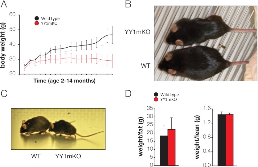
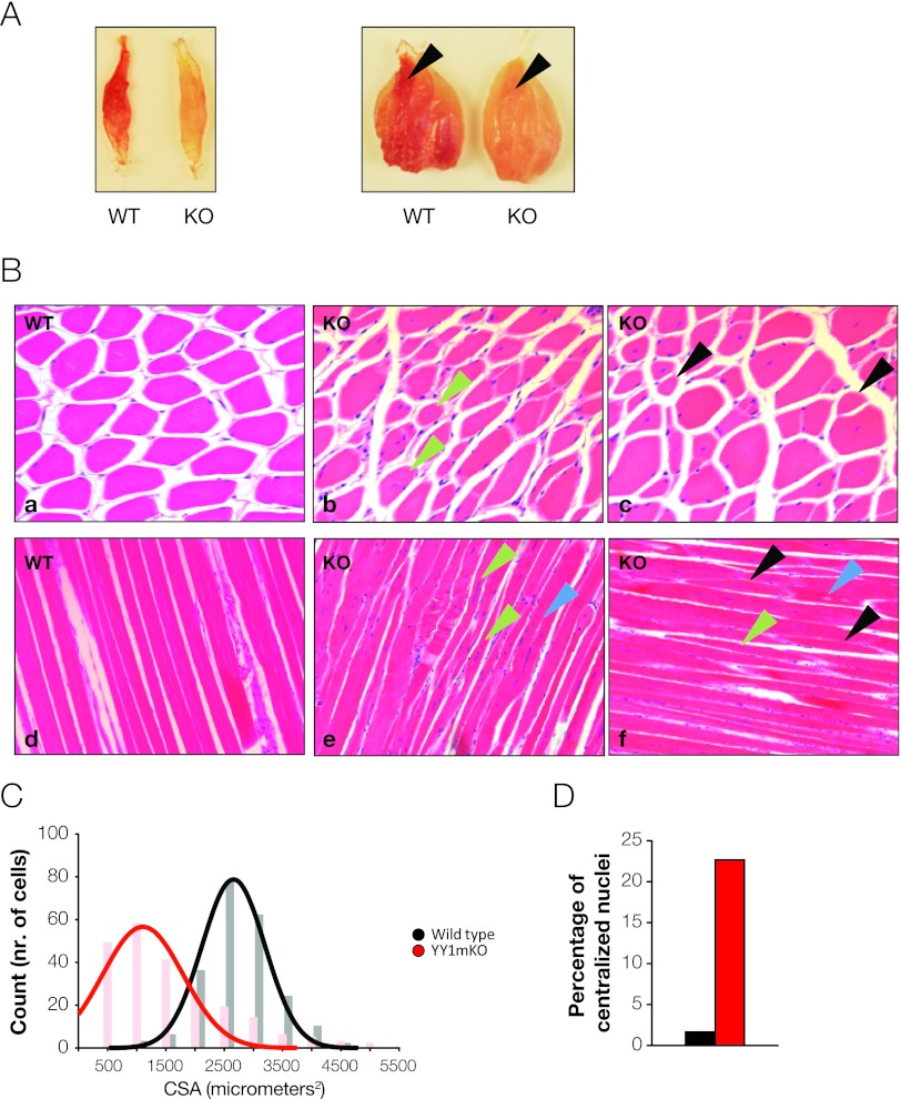
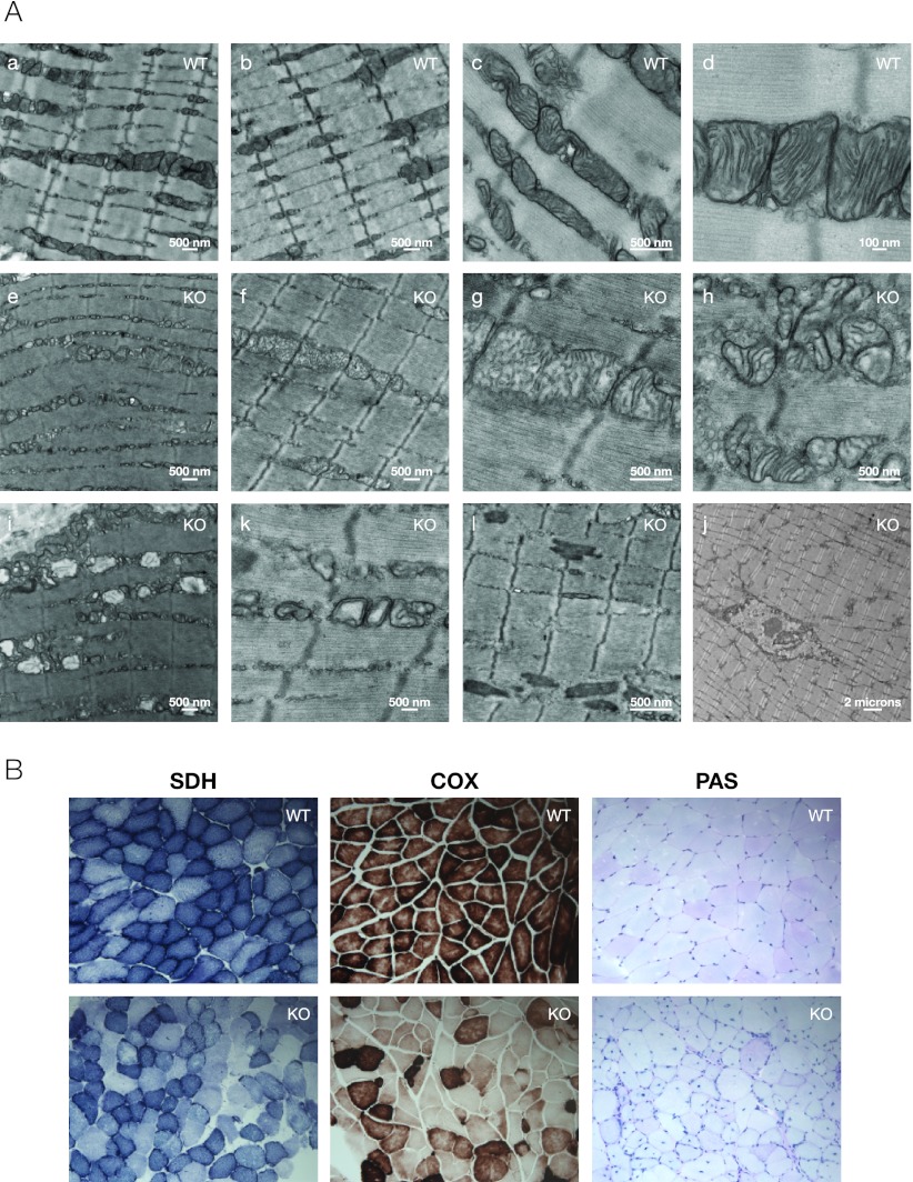
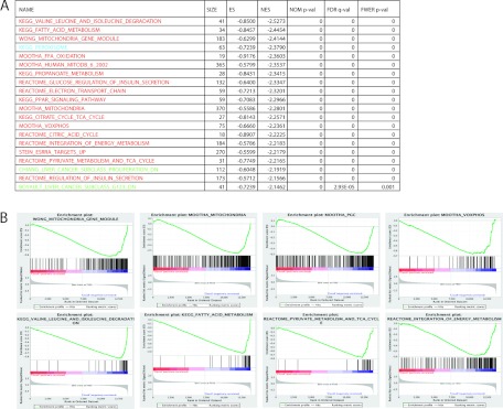
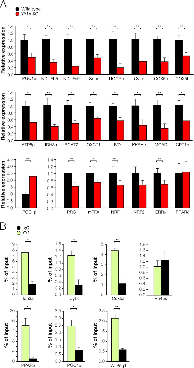
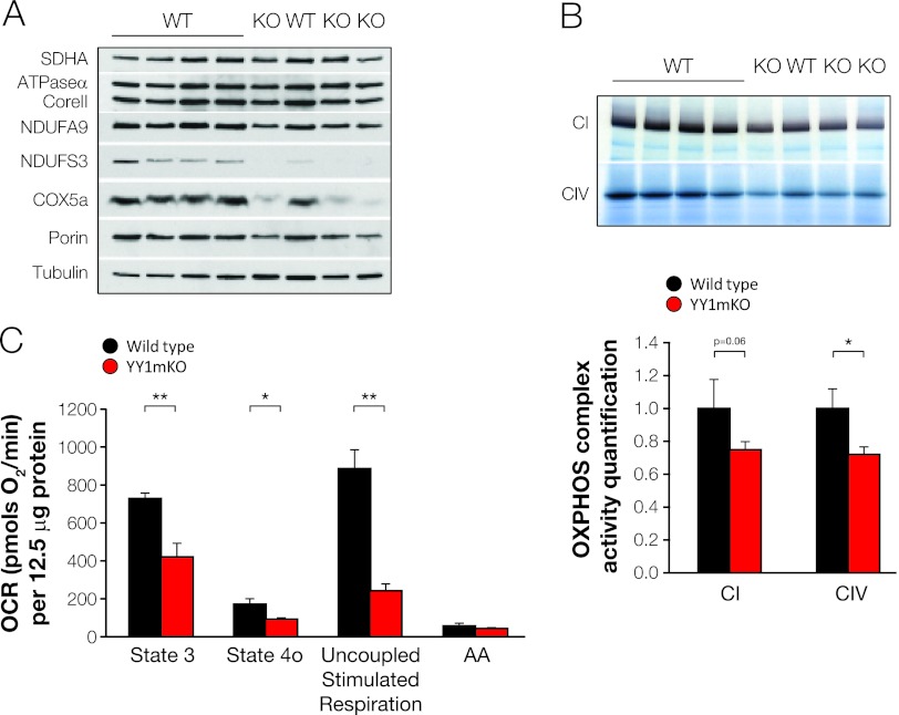
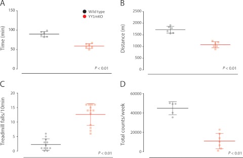
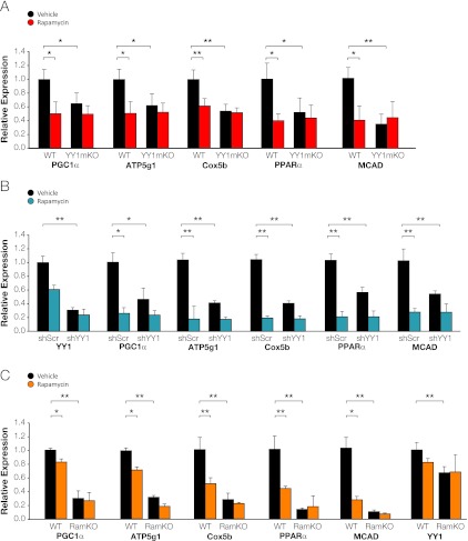
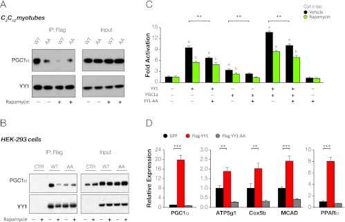
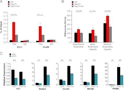
The formation, distribution, and maintenance of functional mitochondria are achieved through dynamic processes that depend strictly on the transcription of nuclear genes encoding mitochondrial proteins. A large number of these mitochondrial genes contain binding sites for the transcription factor Yin Yang 1 (YY1) in their proximal promoters, but the physiological relevance is unknown. We report here that skeletal-muscle-specific YY1 knockout (YY1mKO) mice have severely defective mitochondrial morphology and oxidative function associated with exercise intolerance, signs of mitochondrial myopathy, and short stature. Gene set enrichment analysis (GSEA) revealed that the top pathways downregulated in YY1mKO mice were assigned to key metabolic and regulatory mitochondrial genes. This analysis was consistent with a profound decrease in the level of mitochondrial proteins and oxidative phosphorylation (OXPHOS) bioenergetic function in these mice. In contrast to the finding for wild-type mice, inactivation of the mammalian target of rapamycin (mTOR) did not suppress mitochondrial genes in YY1mKO mice. Mechanistically, mTOR-dependent phosphorylation of YY1 resulted in a strong interaction between YY1 and the transcriptional coactivator peroxisome proliferator-activated receptor gamma coactivator 1α (PGC1α), a major regulator of mitochondrial function. These results underscore the important role of YY1 in the maintenance of mitochondrial function and explain how its inactivation might contribute to exercise intolerance and mitochondrial myopathies.


Figures

Fig 1
Fig 1
Specific genetic deletion of YY1 in skeletal muscle results in a dwarf phenotype. (A) The body weight of wild-type (WT) or YY1mKO mice (n, 9 to 10) was monitored for 1 year from the age of 2 months. (B) YY1mKO mice are smaller than wild-type mice (age, 1 year). (C) YY1mKO mice develop kyphosis (age, 1 year). (D) MRI of YY1mKO and wild-type mice (age, 1 year; n = 10).
Fig 2
Fig 2
Deficiency of YY1 in skeletal muscle results in morphological mitochondrial defects. (A) Appearance of skeletal muscles of 3-month-old wild-type (WT) and YY1mKO mice. (Left) Soleus muscle; (right) the whole calf (gastrocnemius, soleus, and plantaris muscles). Arrowheads indicate the soleus. (B) (Top) Cross sections of gastrocnemius from 6-month-old male wild-type (a) or YY1mKO (b and c) mice stained with H&E. (Bottom) Longitudinal sections from wild-type (d) or YY1mKO (e and f) mice stained with H&E. Black arrowheads indicate small fibers; green arrowheads point to cells with centralized nuclei, and blue arrowheads point to central core-like structures. (C) Cross-sectional area (CSA) distribution in 6-month-old wild-type or YY1mKO fibers. (D) Percentages of cells with centralized nuclei in 6-month-old wild-type or YY1mKO fibers.
Fig 3
Fig 3
(A) Representative electron micrographs of soleus muscles of 6-month-old male wild-type (WT) (a to d) and YY1mKO (e to l) mice. (B) Histochemistry of mitochondrial proteins and glycogen. Succinate dehydrogenase (SDH) and cytochrome oxidase (COX) activities were determined, and glycogen staining with periodic acid-Schiff stain (PAS) was performed, on sections of gastrocnemius muscle from 6-month-old male mice.
Fig 4
Fig 4
Mitochondrial metabolic genes are downregulated in YY1mKO skeletal muscle. (A) GSEA of the top 20 pathways downregulated in soleus muscles of 3-month-old YY1mKO mice. Red represents mitochondrial pathways, green represents cancer-related pathways, and blue represents the peroxisomal pathway. ES, enrichment score; NES, normalized enrichment score; NOM p-val, nominal P value; FDR q-val, probability for false discovery rate; FWER p-val, familywise error rate. (B) Enrichment plots for representative gene expression sets. The color bar depicts the gene list used in the GSEA ordered by differential gene expression. Red indicates higher (positively correlated) and blue indicates lower(negatively correlated) signal to noise in WT compared to YY1mKO mice.
Fig 5
Fig 5
Deficiency of YY1 in skeletal muscle results in decreases in the expression of mitochondrial and transcriptional regulatory mitochondrial genes. (A) Gene expression was measured by quantitative real-time PCR from soleus muscle RNA extracted from 3 month-old fed mice. (B) ChIP analysis from gastrocnemius muscles of 3 month-old fed mice. All values are presented as means ± SD. Four to 10 mice were used. *, P < 0.05; **, P < 0.01.
Fig 6
Fig 6
YY1mKO skeletal muscles have lower expression of mitochondrial proteins and lower levels of mitochondrial electron chain respiratory activity. (A) Western blots of mitochondrial proteins in skeletal muscle. (B) OXPHOS complex activities measured by in gel-activity assays (top) and relative quantification (bottom). (C) Oxygen consumption rates (OCR) measured from total mitochondrial fractions isolated from wild-type and YY1mKO gastrocnemius muscles. Bars show average OCR values from 4 to 5 mice per group ± standard errors of the means. The substrates used were 5 mM pyruvate plus 5 mM malate. State 3 (OCR associated with maximal ATP synthesis rates) was induced with 1 mM ADP. State 4o (proton leak, respiration independent of ATP synthesis) was induced by 2 μM oligomycin (complex V inhibitor). Electron transport chain activity independent of ATP synthesis (uncoupled) was induced by 4 μM FCCP. Nonmitochondrial electron transport OCR (background) were determined by the addition of the complex III inhibitor antimycin A (AA). Asterisks indicate significant differences (*, P < 0.05; **, P < 0.01) by an unpaired, two-tailed Student t test.
Fig 7
Fig 7
YY1mKO mice exhibit exercise intolerance. (A and B) Six-month-old male wild-type and YY1mKO mice were subjected to forced treadmill performance until exhaustion, and time (A) and distance (B) were recorded. (C) Number of stops/falls from the treadmill. (D) Voluntary wheel performance. Five to 13 mice were tested. *, P < 0.01.
Fig 8
Fig 8
Rapamycin does not suppress mitochondrial genes in YY1mKO or RamKO mice. (A) Six-month-old male wild-type and YY1mKO mice were treated with a vehicle or 2.5 mg/kg rapamycin for 14 days. Gene expression was measured by quantitative real-time PCR from soleus RNA extracted from fed mice. (B) C2C12 myotubes were infected with control scrambled shRNA (shScr) or shYY1 for 48 h, and gene expression was measured by quantitative real-time PCR. (C) Three-month-old male wild-type or RamKO mice were treated with a vehicle or 2.5 mg/kg rapamycin for 14 days. Gene expression was measured by quantitative real-time PCR from soleus RNA extracted from fed mice. All values are presented as means ± SD. Four to 10 mice were used. *, P < 0.05; **, P < 0.01.
Fig 9
Fig 9
mTORC1-dependent phosphorylation of YY1 recruits PGC1α and increases mitochondrial gene expression and bioenergetic function. (A) Coimmunoprecipitation of Flag-labeled YY1 and hemagglutinin (HA)-labeled PGC1α in C2C12 myotubes treated with a vehicle or 20 nM rapamycin for 2 h. IP, immunoprecipitation. (B) Coimmunoprecipitation of Flag-YY1 and HA-PGC1α in HEK-293 cells treated with a vehicle or 20 nM rapamycin for 2 h. (C) Luciferase assay in HEK-293 cells transfected with a cytochrome c luciferase construct and the indicated proteins. Rapamycin (20 nM) was added as indicated 24 h before cell lysis. Data are expressed as fold activation and were normalized to the level of expression in an empty-vector-transfected control. a, significant difference (P < 0.01) between the control and the overexpressed plasmid; b, significant difference (P < 0.01) between the vehicle and rapamycin; **, P < 0.01. (D) Primary muscle myotubes were infected with green fluorescent protein (GFP), Flag-YY1, or Flag-YY1-AA for 48 h, and gene expression was measured by quantitative real-time PCR. All values are presented as means ± SD. Four to 10 mice were used. **, P < 0.01; ***, P < 0.001.
Fig 10
Fig 10
YY1 binds to nuclear mitochondrial genes and increases oxygen consumption in cultured muscle cells. (A) ChIP was performed in C2C12 myotubes using antibodies specific for Flag or IgG. All values are presented as means ± standard errors of the means. Four mice were used. *, P < 0.05; **, P < 0.01; ***, P < 0.001. Cyt c, cytochrome c. (B) Oxygen consumption rates (OCR) in C2C12 myotubes infected with green fluorescent protein (GFP), Flag-YY1, or Flag-YY1-AA. Mitoch, mitochondrial. (C) C2C12 myotubes were infected with either shScrambled or shYY1 and with GFP or PGC1α for 72 h, and gene expression was measured by quantitative real-time PCR. All values are presented as means ± SD. Four to 10 mice were used. *, P < 0.05; **, P < 0.01.

Similar articles

Cited by

References

    1. Affar EB, et al. 2006. Essential dosage-dependent functions of the transcription factor yin yang 1 in late embryonic development and cell cycle progression. Mol. Cell. Biol. 26:3565–3581 - PMC - PubMed
    1. Atchison L, Ghias A, Wilkinson F, Bonini N, Atchison ML. 2003. Transcription factor YY1 functions as a PcG protein in vivo. EMBO J. 22:1347–1358 - PMC - PubMed
    1. Austen M, Luscher B, Luscher-Firzlaff JM. 1997. Characterization of the transcriptional regulator YY1. The bipartite transactivation domain is independent of interaction with the TATA box-binding protein, transcription factor IIB, TAFII55, or cAMP-responsive element-binding protein (CPB)-binding protein. J. Biol. Chem. 272:1709–1717 - PubMed
    1. Bentzinger CF, et al. 2008. Skeletal muscle-specific ablation of raptor, but not of rictor, causes metabolic changes and results in muscle dystrophy. Cell Metab. 8:411–424 - PubMed
    1. Blättler SM, et al. 2012. Yin Yang 1 deficiency in skeletal muscle protects against rapamycin-induced diabetic-like symptoms through activation of insulin/IGF signaling. Cell Metab. 15:505–517 - PMC - PubMed

Publication types