. 2012 Aug;32(16):3321-32.
doi: 10.1128/MCB.00296-12. Epub 2012 Jun 18.

Calpain 2 activated through N-methyl-D-aspartic acid receptor signaling cleaves CPEB3 and abrogates CPEB3-repressed translation in neurons

Affiliations

Calpain 2 activated through N-methyl-D-aspartic acid receptor signaling cleaves CPEB3 and abrogates CPEB3-repressed translation in neurons

Chia-Fang Wang et al. Mol Cell Biol. 2012 Aug.

Abstract

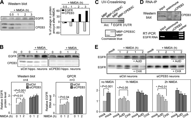
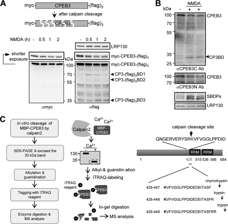
Long-term memory requires the activity-dependent reorganization of the synaptic proteome to modulate synaptic efficacy and consequently consolidate memory. Activity-regulated RNA translation can change the protein composition at the stimulated synapse. Cytoplasmic polyadenylation element-binding protein 3 (CPEB3) is a sequence-specific RNA-binding protein that represses translation of its target mRNAs in neurons, while activation of N-methyl-d-aspartic acid (NMDA) receptors alleviates this repression. Although recent research has revealed the mechanism of CPEB3-inhibited translation, how NMDA receptor signaling modulates the translational activity of CPEB3 remains unclear. This study shows that the repressor CPEB3 is degraded in NMDA-stimulated neurons and that the degradation of CPEB3 is accompanied by the elevated expression of CPEB3's target, epidermal growth factor receptor (EGFR), mostly at the translational level. Using pharmacological and knockdown approaches, we have identified that calpain 2, activated by the influx of calcium through NMDA receptors, proteolyzes the N-terminal repression motif but not the C-terminal RNA-binding domain of CPEB3. As a result, the calpain 2-cleaved CPEB3 fragment binds to RNA but fails to repress translation. Therefore, the cleavage of CPEB3 by NMDA-activated calpain 2 accounts for the activity-related translation of CPEB3-targeted RNAs.


Figures

Fig 1
Fig 1
NMDA-induced EGFR expression is mainly through CPEB3-controlled translation. (A) The Western blots of EGFR and CPEB3 using hippocampal neurons treated with 50 μM NMDA (+ NMDA) or not treated with NMDA for the indicated times (in hours). The changes in EGFR and CPEB3 protein levels after NMDA treatment are shown as a percentage of change compared to the signal obtained in unstimulated neurons. (B) The EGFR protein and RNA levels in control (siCtrl) and CPEB3 knockdown (siCPEB3) neurons treated with NMDA or not treated with NMDA for the indicated time were detected by Western blotting and RT-QPCR assay, respectively. The bar graphs at the bottom of the panel display the relative EGFR protein and RNA levels under different time treatments of NMDA with the signal from nontreated siCtrl neurons set at 1. The results were analyzed and expressed as means plus SEMs (error bars). An asterisk marks a statistical difference in the EGFR protein and RNA levels (two-tailed Student's t test) compared to those of nontreated cells. (C) The recombinant maltose-binding protein (MBP) and MBP fused to the RNA-binding domain of CPEB3 (MBP-CPEB3C) were UV cross-linked with 32P-labeled 3′-UTRs of Arc and EGFR RNAs, RNase A treated, and then analyzed by SDS-PAGE. The bottom panel shows Coomassie blue-stained proteins. (D) RNA immunoprecipitation (RNA-IP) using cortical neuronal lysate incubated with preimmune or CPEB3 IgG. EGFR RNA in the precipitated substances was detected by RT-PCR. αCPEB3, anti-CPEB3 antibody. (E) The Western blots of EGFR and CPEB3 using control (siCtrl) and CPEB3 knockdown (siCPEB3) neurons treated with NMDA or not treated with NMDA in the presence (+) or absence of 2 μg/ml actinomycin (ActD) or 50 μg/ml cycloheximide (CHX). The bar graph displays the relative EGFR protein level under different treatments with the signal from nontreated siCtrl neurons set at 1. The results from four independent experiments were analyzed and expressed as means plus SEMs. An asterisk denotes a statistical difference in the EGFR protein level (two-tailed Student's t test) compared to that of mock-treated cells.
Fig 2
Fig 2
Blockade of calpain activity inhibits NMDA-induced CPEB3 degradation. (A and B) The cortical neurons at DIV 11 or 12 were treated with 50 μM NMDA for 3 min and then harvested at the indicated times for Western blotting with CPEB3 and leucine-rich PPR motif-containing protein (LRP130) antibodies (A) or for RT-QPCR to measure the relative CPEB3 RNA level (B). (C) Similar to panel A but the lysates were also used for Western blotting of CPEB3, EGFR, and LRP130. The results from 4 independent experiments were analyzed and expressed as means plus SEMs. An asterisk marks a statistical difference in CPEB3 and EGFR protein levels with or without 3 min of stimulation of NMDA (two-tailed Student's t test). (D) The cortical neurons were pretreated without (mock) or with 20 μM MG132 or 10 μM lactacystin for 15 min prior to the presence (+) or absence (−) of NMDA stimulation (3-min pulse). The neurons were harvested 2 h after for Western blot analysis. (E and F) Similar to panel D, except 20 μM calpain inhibitor III and 2 mM EGTA were used to treat neurons before the pulse stimulation of NMDA.
Fig 3
Fig 3
NMDA-induced CPEB3 degradation is ameliorated in the Capns1 knockdown neurons. (A) Schematic diagram showing the domain structure of the catalytic subunits of calpain 1 and calpain 2, CAPN1 and CAPN2, respectively. The small regulatory subunit, Capns1, binds to the domain IV of CAPN. The catalytic triad, Cys-His-Asn, located in domain II, is essential for protease activity. a.a., amino acids. (B) The two shRNAs, siCapns1#1 and siCapns1#2, targeted against the mouse Capns1 were introduced into mouse cortical neurons using lentiviral infection. The Capns1 mRNA level in control (siCtrl) and knockdown (siCapns1) neurons was analyzed by RT-QPCR. (C) The siCtrl and siCapns1 neurons were treated with a 3-min pulse of 50 μM NMDA or not treated with NMDA and then collected 1 or 2 h later for Western blot analysis using CPEB3, LRP130, and αII-spectrin (SBDPs, αII-spectrin breakdown products) antibodies. The results were analyzed and expressed as means plus SEMs. An asterisk marks a statistical difference in the CPEB3 protein level (two-tailed Student's t test).
Fig 4
Fig 4
Calpain 2 proteolyzes CPEB3 in the NMDA-treated neurons. (A) The two shRNAs, siCAPN1#1 (or siCAPN2#1) and siCAPN1#2 (or siCAPN2#2), targeted against the rat CAPN1 (or CAPN2) were introduced into rat neurons using lentiviral infection. The CAPN1 and CAPN2 RNA levels in the siCtrl, siCAPN1, and siCAPN2 neurons were detected by RT-QPCR from two independent measurements. The results were expressed as means plus SEMs. (B) The control and various knockdown neurons treated with a 3-min pulse of 50 μM NMDA or not treated with NMDA were analyzed 2 h later by Western blotting. The results were expressed as means plus SEMs. An asterisk denotes a significant difference in the CPEB3 protein level (two-tailed Student's t test). (C) The cultured cortical neurons were treated with 20 μM calpain inhibitor III along with NMDA stimulation or without NMDA stimulation. The cell lysates were pulled down with control or CPEB3 IgG and probed with CAPN2 and CPEB3 antibodies. IP, immunoprecipitation; IB, immunoblotting. (D) The top diagram denotes the various CPEB3 truncated mutants. The 293T cell lysates containing Flag-tagged CAPN2 along with myc-tagged full-length (wild-type [wt]) or truncated mutant CPEB3 were precipitated with the myc antibody and immunoprobed with myc and Flag antibodies. RRM, RNA recognition motif; Zif, zinc finger.
Fig 5
Fig 5
CPEB3 is degraded by NMDA-activated calpain 2 in the cytoplasmic and synaptic compartments. (A) Cultured hippocampal neurons of DIV 12 or 13 were treated with 50 μM NMDA for 3 min or not treated with NMDA and immunostained 30 min later with CAPN2 and CPEB3 antibodies. (B) The DIV 12 cortical neurons were treated with 50 μM NMDA for 3 min or not treated with NMDA and fractionated for cytosol and nuclear extracts at the indicated times (20 min or 1 or 2 h). The lysates were used for immunoblotting. (C) Low- and high-magnification views of CAPN2 and CPEB3 images are shown. The white arrowheads mark colocalized CAPN2 and CPEB3 in the dendrite. Bars, 20 μm (top) and 5 μm (bottom). (D) The postnuclear supernatant prepared from the rat brain was centrifuged at 12,000 × g (12k xg), and the supernatant (sup) and synaptosome-enriched pellet are shown below the blot. The synaptosomes were aliquoted and stimulated with 50 μM NMDA for the indicated times and harvested for Western blotting of CPEB3 and synaptophysin (sy38). The results from 5 independent synaptosome preparations were analyzed and expressed as means plus SEMs. An asterisk denotes a significant difference in the CPEB3 protein level (two-tailed Student's t test).
Fig 6
Fig 6
CPEB3 is cleaved by calpain 1 and calpain 2 in vitro. (A) The [35S]Met/Cys-radiolabeled p53 and CPEB3 were synthesized using the reticulocyte lysate system and then mixed with the Jurkat cell cytoplasmic extract in the presence or absence of 0.2 mM CaCl2 along with other indicated chemicals (EGTA, dimethyl sulfoxide [DMSO], and calpain inhibitor III [Cal inh III]) at 37°C for 1 h. The radioactive mixtures were separated on SDS-polyacrylamide gels and analyzed by a phosphorimager. The arrowheads indicate the positions of major breakdown fragments after cleavage. (B) The recombinant maltose-binding protein (MBP)–CPEB3 was incubated with or without purified calpain 1 or calpain 2 in the Ca2+-containing buffer along with other indicated reagents at 37°C for 1 h. The mixtures were separated by SDS-PAGE and stained with Coomassie blue. The asterisks denote various breakdown products, CAPN1BD, CAPN2 BD, and CP3BD, derived from CAPN1, CAPN2, and CPEB3, respectively.
Fig 7
Fig 7
Identification of the calpain cleavage site at the extreme C terminus of CPEB3. (A) The neurons infected with the lentivirus expressing myc-CPEB3-(Flag)3 were treated with NMDA and harvested at the indicated times. The neuronal lysates were immunoblotted with LRP130, myc, and Flag antibodies. The arrowheads show the positions of three breakdown fragments, CP3-(Flag)3BD1, -BD2, and -BD3. (B) The NMDA-treated neuronal lysates were analyzed by Western blotting using antibodies recognizing the N or C terminus of CPEB3 (anti-CPEB3N antibody [αCPEB3N Ab] or anti-CPEB3C), SBDPs, and LRP130. (C) Work flow used to determine the cleavage site on CPEB3. The calpain 2 cleaved C-terminal fragment of MBP-CPEB3 separated by SDS-PAGE was isolated and alkylated, guanidinated, and then labeled with the iTRAQ reagent on the gel. The labeled gel was digested with trypsin or chymotrypsin, which cleaved bonds at the carboxyl sides of lysine/arginine residues or tyrosine/tryptophan/phenylalanine residues, respectively. The collected peptides were analyzed by tandem mass spectrometry. The iTRAQ-conjugated peptides are listed on the right.
Fig 8
Fig 8
The cleavage of CPEB3 by calpain 2 abolishes its translational repression activity. (A) The recombinant MBP or MBP-CPEB3 was incubated with calpain 2 in the absence or presence of Ca2+, the mixtures were incubated with the 32P-labeled 1904 RNA probe. One half of the RNA-protein mixture was used for the gel retardation assay (top gel). The other half was UV cross-linked and RNase A digested, followed by SDS-PAGE separation. The gel was stained with Coomassie blue and exposed to a phosphorimager. The RNA-protein complexes are labeled with one or two asterisks. (B) RNA reporter assay. The hippocampal or cortical neurons of DIV 10 or 11 were transfected with mRNAs encoding β-galactosidase (β-gal), myc-CPEB3 or myc-CP3BD, along with the firefly luciferase appended to the EGFR 3′-UTR and Renilla luciferase. (C) Similar to panel B, the transfected neurons were stimulated with 50 μM NMDA or not stimulated with NMDA. The neurons were harvested 3 h later for Western blotting and luciferase assays. The normalized luciferase activity (firefly/Renilla) was calculated. The results were analyzed and expressed as means plus SEMs. An asterisk marks a statistical difference (two-tailed Student's t test). The intensities of immunodetected signals analyzed by the ImageJ software are displayed as relative ratios at the bottoms of blots. (D) Schematic model of CPEB3-governed translation. The binding of CPEB3 to the 3′-UTR of plasticity-related protein (PRP) RNA, such as EGFR, reduces the translation through its interaction with eEF2. NMDA-induced EGFR RNA translation is in part caused by the calpain 2-dependent cleavage of CPEB3, which presumably allows eEF2 to resume its maximal GTPase activity and enhances the translation of EGFR RNA.

Similar articles

Cited by

References

    1. Abe K, Takeichi M. 2007. NMDA-receptor activation induces calpain-mediated beta-catenin cleavages for triggering gene expression. Neuron 53:387–397 - PubMed
    1. Adamec E, Beermann ML, Nixon RA. 1998. Calpain I activation in rat hippocampal neurons in culture is NMDA receptor selective and not essential for excitotoxic cell death. Brain Res. Mol. Brain Res. 54:35–48 - PubMed
    1. Arthur JS, Elce JS, Hegadorn C, Williams K, Greer PA. 2000. Disruption of the murine calpain small subunit gene, Capn4: calpain is essential for embryonic development but not for cell growth and division. Mol. Cell. Biol. 20:4474–4481 - PMC - PubMed
    1. Ashby MC, et al. 2004. Removal of AMPA receptors (AMPARs) from synapses is preceded by transient endocytosis of extrasynaptic AMPARs. J. Neurosci. 24:5172–5176 - PMC - PubMed
    1. Banker G, Goslin K. 1988. Developments in neuronal cell culture. Nature 336:185–186 - PubMed

Publication types