. 2012 Mar 1;2(1):15-30.
doi: 10.1016/j.baga.2012.01.002. Epub 2012 Feb 11.

Anatomy of Graft-induced Dyskinesias: Circuit Remodeling in the Parkinsonian Striatum

Affiliations

Anatomy of Graft-induced Dyskinesias: Circuit Remodeling in the Parkinsonian Striatum

Kathy Steece-Collier et al. Basal Ganglia. .

Abstract

The goal of researchers and clinicians interested in re-instituting cell based therapies for PD is to develop an effective and safe surgical approach to replace dopamine (DA) in individuals suffering from Parkinson's disease (PD). Worldwide clinical trials involving transplantation of embryonic DA neurons into individuals with PD have been discontinued because of the often devastating post-surgical side-effect known as graft-induced dyskinesia (GID). There have been many review articles published in recent years on this subject. There has been a tendency to promote single factors in the cause of GID. In this review, we contrast the pros and cons of multiple factors that have been suggested from clinical and/or preclinical observations, as well as novel factors not yet studied that may be involved with GID. It is our intention to provide a platform that might be instrumental in examining how individual factors that correlate with GID and/or striatal pathology might interact to give rise to dysfunctional circuit remodeling and aberrant motor output.


Figures

Figure 1
Figure 1. Differential time course of expression of levodopa-induced dyskinesias (LIDs) versus graft-induced dyskinesias (GID)
The arrow demarks the time of graft surgery. As the graft matures, the incidence of LID lessens and there is an emergence of GID behaviors, e.g.: [9,10,17,20,21].
Figure 2
Figure 2. Modeling Graft-induced Dyskinesia: Does Graft Size Matter?
This diagram is used to consider whether it is it possible that the unique post-graft dyskinetic behaviors noted in either parkinsonian rats or humans are differentially responsive to dopamine replacement therapy by virtue of graft size. In considering this, it is noteworthy that in the Denver/Columbia clinical trial after transplantation, the increase in overall putamenal PET 18F-DOPA signal in the GID-expressing patient group was twice that of the GID-negative group at 12 months (P < 0.03) and almost three times larger at 24 months (P < 0.005) [27]. Despite the 18F-DOPA signal being 3 times higher in the GID expressing patients, it was still below the level of the normal striatum by about 20% [27]. It is interesting to speculate whether the GID-expressing patient group could be represented by Graft ‘X’ and the GID-negative group represented by Graft ‘Y’ (both Graft ‘X’ and ‘Y’ appear in the right hand panel of graph). If one compares the amount of tissue that has been grafted into the parkinsonian rat in models examining GID-like behavior, there is substantially less tissue proportionately grafted in the rodent models compared to most human trials. Rat Model: [20]: 1 ventral mesencephalon (VM) with postmortem TH+ cell number = 2,800; [21]: < 1 VM with postmortem TH+ cell number = 280 (small grafts) or 17,408 (large grafts). Human Grafting: [9]: noodles; postmortem TH+ cell number = 62,507; [10]: 1–4 VM per patient with 70,000–120,000 TH+ cells per side; [12,13]: 6 VM per patient; postmortem TH+ cell number= 210,067; [17]: 6.3 ± 2.8 VM per patient, TH+ number not available. Thus, is it possible that the smaller number of grafted DA neurons in the rat brain requires a DA agonist to “push” the levels of DA into the biphasic dyskinesia range to elicit expression of GID? The case presented here is undoubtedly an oversimplification in that it does not account for potential differences in DA neuron biology between rodents and humans, the degree of DA neuron survival or neurite outgrowth, and the relative striatal volume influenced by grafts in the two situations. If one just looks at the number of DA neurons grafted per volume of the striatum between rat and human, such calculations remain equivocal in answering this question. For example, the human putamen (common clinical grafting site) can be estimated to be approximately 100× larger than the rat striatum [–, –175], and there are approximately 10× more DA neurons in the human SN compared to the rat SN [–, –173]. Based on such estimates grafting 1 ventral mesencephalon into a rat striatum would result in approximately 10,000 DA cells /0.06 cm3 or 166,666 DA cells/cm3, which was the paradigm done by [20, 21] and one in which LD or amphetamine is apparently necessary to “push” striatal DA to levels that induce reliable experimental GID. In contrast, grafting 6 VM into the human striatum/putamen as was done by [17] would result in approximately 600,000 DA cells/ 6.25 cm3 or 96,000 DA cells/cm3, a scenario where clinical GID are expressed “OFF” LD. In these two example comparisons the density of DA neurons/cm3 in the human striatum/putamen would be approximately 60% of that found in the rat striatum (assuming equal degree of DA neuron survival). However, this comparison would be accurate only if the grafted cells spread uniformly through the entire “striatum”. It is well documented that grafted DA neurons remain largely confined within and immediately surrounding the injection site [e.g.; 20,21,10,13], with the bolus of grafted neurons giving rise to neurites that extend into the target striatum, varying in density and distance. Thus, factors including neurite outgrowth and degree of regional DA replacement need to be considered when pondering the question above, which is suggested by the behavioral and pharmacological differences associated with the model and the patient. Abbreviations: 1) “ON” and “OFF” refer to the relationship with the intake of levodopa medication, i.e.; on-medication and off-medication. 2) The Y-axis label “Plasma LD/Striatal DA” refers to the plasma concentration of levodopa (LD) and the likely corresponding striatal concentration of DA, with lowest levels represented at the X–Y axis intercept, and higher levels at the top of the Y-axis. 3) +LD or +LD/amphet refers to administration of the drug LD or amphetamine to grafted subjects.
Figure 3
Figure 3. Schematic Depiction of the Connectivity in the (a) Healthy, (b) Parkinsonian and (c) Dopamine-grafted Parkinsonian Striatum
These diagrams are oversimplifications of particular aspects of striatal circuitry that may be relevant to understanding GID behaviors. Studies on which these diagrams are based are detailed in the body of this review. Numbers is parenthesis ‘()’ in the legend refer to numbers in the figures; numbers in brackets ‘[ ]’ refer to citations. (a) In the healthy striatum, descending glutamate fibers (purple) from the cerebral cortex and thalamus make asymmetric contacts (2) preferentially onto the head of numerous dendritic spines (1) of the resident MSNs. This input is modulated by ascending DA fibers (blue) from the substantia nigra pars compacta (3) that make symmetric en passant contacts preferentially onto the necks of those same spines. Additionally there are a small number of 5HT fibers from the raphe nucleus (not depicted) that project to the striatum, although these typically do not form synapses with the MSNs. (b) In the parkinsonian striatum, following severe dopamine depletion, there is a significant loss of dendritic spines (1) of the MSNs of the indirect pathway in rodents [56,57], and direct and indirect pathways in non-human primates [58]. With the loss of these critical input sites both glutamatergic (purple) (2) and remaining DAergic (blue) (3) fibers show an increase in the number of atypical contacts onto dendrites rather than onto their normal spinous targets [19]. Additionally, there is an increase in the number of ectopic serotonergic fibers (orange) (4) [e.g.: 34]; and in parkinsonian rats treated with dyskinogenic levodopa there is a larger proportion of these making actual synaptic contacts onto the MSN (not depicted) [e.g.: 34]. Finally, there is an increase in inflammation in the parkinsonian striatum depicted here with the presence of activated microglia (yellow) (5). (c) In the dopamine-grafted parkinsonian striatum, despite some restoration of DAergic tone, current data suggests that there remains a significant loss of dendritic spines (1) on MSNs. In addition to making atypical contacts onto dendrites, glutamate (purple) (2) and DA (blue) (3) fibers show additional changes in their specialization and symmetry, with glutamate fibers making more perforated contacts (2) [19], which is generally associated with synaptic plasticity, and DA fibers making more asymmetrical contacts (3), which are atypical for DA fibers, and is classically associated with excitatory synapses. There is also presence of excitatory synapses onto the grafted cells themselves (6) [19]. There continues to be an increase in the presence of 5HT fibers (orange), potentially from both the raphe nucleus as well as from serotonergic cells within the grafts themselves (4). Since all grafted PD patients to date have undergone long-term levodopa treatment, there should be a marked increased incidence of synapses (darkened orange lines) between these ectopic 5HT fibers and MSNs [34]. Additionally, pre-synaptic vesicles in 5HT terminal boutons co-express vGLUT3 protein (7) [–44], indicative of co-transmission of glutamate at these monoaminergic synapses. Similarly, following striatal DA depletion in the rat, there is re-expression of vGLUT2 protein (7) [68]; however, whether this occurs in grafted DA neurons is unknown. Research evidence suggests that much of this atypical connectivity may be modulated by the increased inflammatory response observed in the grafted striatum, depicted here to be influenced by activated microglia (yellow) (5). ‘Perforated synapses’ are depicted with the dashed, thickened post-synaptic arcs opposite the glutamate terminals; ‘Asymmetric synapses’ are depicted with thickened post-synaptic arcs opposite glutamate, DA and 5HT terminals. (7) While VMAT2 and vGLUT proteins likely occur in the same synaptic vesicles of monoaminergic neurons the occurrence of glutamate as a minor vesicular transmitter in the DA and 5HT terminals is depicted in this schematic as a single dark glutamate vesicle, indicating its relative minor presence.
Figure 4
Figure 4
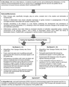
Immune Activation and Graft-induced Dyskinesias. Footnote; Model A: Experimental GID occurred only in dopamine-grafted rats, and not in cell-free, sham-grafted rats, despite the presence of equal numbers of MHC class II+ cells in sham and G21 rats, suggesting that the impact of host immune factors is more likely to be related to graft–host synaptic organization than to a direct action on host signaling pathways.

References

    1. Dunn EH. Primary and secondary findings in a series of attempts to transplant cerebral cortex in the albino rat. J Comp Neurol. 1917;27:565–582.
    1. Lindvall O, Brundin P, Widner H, Rehncrona S, Gustavii B, Frackowiak R, et al. Grafts of fetal dopamine neurons survive and improve motor function in Parkinson’s disease. Science. 1990;247:574–577. - PubMed
    1. Freed CR, Breeze RE, Rosenberg NL, Schneck SA, Kriek E, Qi JX, et al. Survival of implanted fetal dopamine cells and neurologic improvement 12 to 46 months after transplantation for Parkinson’s disease. N Engl J Med. 1992;327:1549–1555. - PubMed
    1. Lindvall O, Sawle G, Widner H, Rothwell JC, Björklund A, Brooks D, et al. Evidence for long-term survival and function of dopaminergic grafts in progressive Parkinson’s disease. Ann Neurol. 1994;35:172–180. - PubMed
    1. Peschanski M, Defer G, N’Guyen JP, Ricolfi F, Monfort JC, Remy P, et al. Bilateral motor improvement and alteration of L-dopa effect in two patients with Parkinson’s disease following intrastriatal transplantation of foetal ventral mesencephalon. Brain. 1994;117:487–499. - PubMed

LinkOut - more resources