. 2012 Jun 19:13:15.
doi: 10.1186/1471-2121-13-15.

Identification of novel mitosis regulators through data mining with human centromere/kinetochore proteins as group queries

Affiliations

Identification of novel mitosis regulators through data mining with human centromere/kinetochore proteins as group queries

Aaron R Tipton et al. BMC Cell Biol. .

Abstract

Background: Proteins functioning in the same biological pathway tend to be transcriptionally co-regulated or form protein-protein interactions (PPI). Multiple spatially and temporally regulated events are coordinated during mitosis to achieve faithful chromosome segregation. The molecular players participating in mitosis regulation are still being unravelled experimentally or using in silico methods.

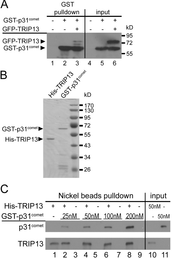
Results: An extensive literature review has led to a compilation of 196 human centromere/kinetochore proteins, all with experimental evidence supporting the subcellular localization. Sixty-four were designated as "core" centromere/kinetochore components based on peak expression and/or well-characterized functions during mitosis. By interrogating and integrating online resources, we have mined for genes/proteins that display transcriptional co-expression or PPI with the core centromere/kinetochore components. Top-ranked hubs in either co-expression or PPI network are not only enriched with known mitosis regulators, but also contain candidates whose mitotic functions are not yet established. Experimental validation found that KIAA1377 is a novel centrosomal protein that also associates with microtubules and midbody; while TRIP13 is a novel kinetochore protein and directly interacts with mitotic checkpoint silencing protein p31(comet).

Conclusions: Transcriptional co-expression and PPI network analyses with known human centromere/kinetochore proteins as a query group help identify novel potential mitosis regulators.


Figures

Figure 1
Figure 1
Interaction between TRIP13 and p31comet. (A) GST-p31comet was expressed either alone or together with GFP-TRIP13 in asynchronous HEK293 cells. The input lysates and GST-pulldowns were probed with anti-GST and GFP antibodies. Untransfected lysates were used as controls. (B) Coomassie blue staining of SDS-PAGE gel loaded with ~0.4 μg of purified recombinant GST-p31comet and His-tagged TRIP13. (C) In vitro binding assays using recombinant His-TRIP13 at 100 nM and GST-p31comet at increasing concentrations. Proteins associated with washed nickel beads were probed with anti-6 × His and anti-GST antibodies.
Figure 2
Figure 2
Characterization of novel kinetochore protein TRIP13. (A) A GFP-TRIP13 transfected HeLa cell in early prometaphase was stained for centromeres (ACA, purple), BUBR1 (red) and DNA (DAPI, blue) to compare with the GFP signals (green). Images shown are maximum projection of a z-series at 1 μm interval. Bar = 10 μm.(B&C) A GFP-TRIP13 transfected prometaphase cell (B) and a cell progressing into metaphase (C) were co-stained with anti-MAD2 antibody and ACA to show GFP-TRIP13 localization only at MAD2 positive kinetochores, such as the one indicated by arrows in (C).(D) A GFP-TRIP13 transfected cell spun onto a coverslip for mitotic chromosome spread preparation was probed with DAPI and anti-HEC1 antibody. (E) A single mitotic chromosome stained with DAPI, anti-GFP and anti-HEC1 antibodies to show kinetochore localization of GFP-TRIP13.
Figure 3
Figure 3
Subcellular localization of GFP-KIAA1377. HeLa cells transfected with GFP-KIAA1377 (green) were fixed and probed with immunofluorescence. Images at single focal planes are shown. Bar = 10 μm. (A&B) Transfected cells finishing cytokinesis were co-stained with anti-α-tubulin (red in A) or anti-Plk1 antibody (purple in B) and counter-stained with DAPI for DNA (blue). (C) A transfected cell expressing higher level of GFP-KIAA1377 (green) was co-stained for α-tubulin (red) and DNA (blue) to indicate co-localization of GFP-KIAA1377 with microtubule structures. (D) GFP-KIAA1377 is localized at centrosomes. Cells at different stages of cell cycle are distinguished based on anti-centrin 2 (red) and DAPI (blue) staining. The GFP signals in different cells were adjusted to the same scale. Enlarged insets show details of boxed areas
Figure 4
Figure 4
GFP-DDX39 is a centrosomal protein. Asynchronous HeLa cells were transfected with GFP-DDX39 (green), fixed and co-stained with DAPI (blue) and anti-γ-tubulin antibody (red). The cell cycle stages were assigned based on the numbers of γ-tubulin foci and DNA morphology. Bar = 10 μm.
Figure 5
Figure 5
Dynamic subcellular localization of GFP-MELK. (A). Distribution of GFP-MELK signals in the cytoplasm and nuclei in G1 and G2 cells. G1 and G2 cells are differentiated based on characteristic staining patterns of CENP-F (red) [82]. Note nuclear signals of GFP-MELK in G2 cells are neither due to overexpression of GFP fusions (compare the G1 and G2 cells in the top row), nor affected by strong CENP-F staining in the Alexa Fluor 555 channel (compare nuclear GFP signals in G2 and late G2 cells in the bottom row). DNA was counterstained with DAPI. Bar = 10 μm. (B) Dynamic cortical localization of GFP-MELK throughout mitosis. Note the GFP signals at the midzone of a late anaphase cell (6). Bar = 10 μm. (C) A 3-D presentation of two GFP-MELK bands at the presumptive cleavage furrow surrounding two groups of separated chromosomes.

References

    1. Pines J, Rieder CL. Re-staging mitosis: a contemporary view of mitotic progression. Nature cell biology. 2001;3(1):E3–E6. - PubMed
    1. Muller GA, Engeland K. The central role of CDE/CHR promoter elements in the regulation of cell cycle-dependent gene transcription. FEBS J. 277(4):877–893. - PubMed
    1. Laoukili J, Kooistra MR, Bras A, Kauw J, Kerkhoven RM, Morrison A, Clevers H, Medema RH. FoxM1 is required for execution of the mitotic programme and chromosome stability. Nature cell biology. 2005;7(2):126–136. - PubMed
    1. Furuno N, den Elzen N, Pines J. Human cyclin A is required for mitosis until mid prophase. The Journal of cell biology. 1999;147(2):295–306. - PMC - PubMed
    1. Musacchio A, Salmon ED. The spindle-assembly checkpoint in space and time. Nat Rev Mol Cell Biol. 2007;8(5):379–393. - PubMed

Publication types

MeSH terms

Substances