. 2012 Oct;103(10):1788-94.
doi: 10.1111/j.1349-7006.2012.02362.x. Epub 2012 Aug 7.

Programmed cell death 6, a novel p53-responsive gene, targets to the nucleus in the apoptotic response to DNA damage

Affiliations

Programmed cell death 6, a novel p53-responsive gene, targets to the nucleus in the apoptotic response to DNA damage

Kazuho Suzuki et al. Cancer Sci. 2012 Oct.

Abstract

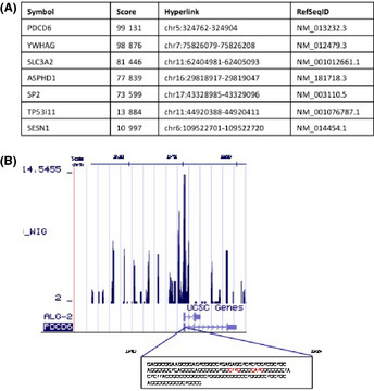
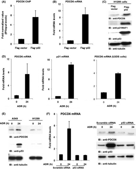
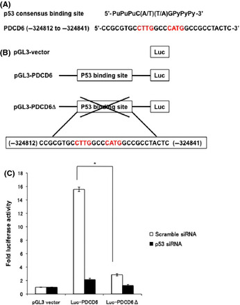
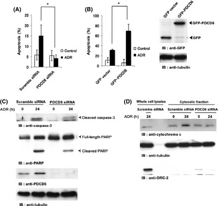
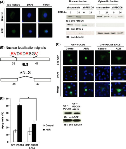
The cellular response to genotoxic stress is multifaceted in nature. Following DNA damage, the tumor suppressor gene p53 activates and plays critical roles in cell cycle arrest, activation of DNA repair and in the event of irreparable damage, induction of apoptosis. The breakdown of apoptosis causes the accumulation of mutant cells. The elucidation of the mechanism for the p53-dependent apoptosis will be crucial in applying the strategy for cancer patients. However, the mechanism of p53-dependent apoptosis remains largely unclear. Here, we carried out ChIP followed by massively parallel DNA sequencing assay (ChIP-seq) to uncover mechanisms of apoptosis. Using ChIP-seq, we identified PDCD6 as a novel p53-responsive gene. We determined putative p53-binding sites that are important for p53 regulation in response to DNA damage in the promoter region of PDCD6. Knockdown of PDCD6 suppressed p53-dependent apoptosis. We also observed that cytochrome c release and the cleavage of PARP by caspase-3 were suppressed by depletion of PDCD6. We further observed that PDCD6 localizes in the nucleus in response to DNA damage. We identified the nuclear localization signal of PDCD6 and, importantly, the nuclear accumulation of PDCD6 significantly induced apoptosis after genotoxic stress. Therefore, we conclude that a novel p53-responsive gene PDCD6 is accumulated in the nucleus and induces apoptosis in response to DNA damage.


Figures

Figure 1
Figure 1
Comprehensive analysis of p53 target gene. (A) The ChIP sequencing score datasets. (B) Peak regions of PDCD6 in sequences obtained from the ChIP sequencing were visualized by the UCSC Genome Browser. The sequence of the highest peak is shown in the box. The p53 binding sites within the sequence of the highest peak are indicated in red color.
Figure 2
Figure 2
PDCD6 is a novel target of p53. (A) PDCD6 expression is regulated by p53. H1299 cells were transfected with Flag vector or Flag‐p53. To perform ChIP assays, choromatin‐protein complexes were immunoprecipitated with anti Flag‐p53 antibodies. Real‐time PCR amplification was performed to analyze the chromatin immunoprecitated fragments using appropriate primers for PDCD6. (B) H1299 cells were transfected with Flag vector or Flag‐p53. The mRNA level of PDCD6 was quantified by real‐time RTPCR. The value is normalized to GAPDH. The data represents the mean ± SD from three independent experiments, each performed in triplicate. (C) H1299 cells were transfected with Flag vector or Flag‐p53 and then analyzed by western blotting. IB, immunoblotting. (D) A549 cells and U2OS cells were treated with 1 µg/mL adriamycin (ADR) and incubated for 24 h. The mRNA level of PDCD6 was quantified by real‐time RTPCR. The value was normalized to GAPDH. The data represent the mean ± SD from three independent experiments, each performed in triplicate. (E) A549 cells and H1299 cells were treated with 1 µg/mL ADR for the indicated times and then analyzed by western blotting. (F) A549 cells were transfected with scramble siRNA or p53 siRNA and treated with 1 µg/mL ADR for 24 h. The mRNA level of PDCD was quantified by real‐time RTPCR. The value was normalized to GAPDH (left). The data represent the mean ± SD from three independent experiments, each performed in triplicate. Cell lysates were subjected to immunoblot analysis with anti‐PDCD6, anti‐p53 or anti‐tubulin (right).
Figure 3
Figure 3
Identification of p53 consensus sequence in PDCD6. (A) The putative p53 response elements are depicted in the upstream of PDCD6. (B) Schematic representation of p53 consensus binding site‐driven luciferase reporter constructs. (C) A549 cells transfected with scramble siRNA or p53 siRNA were transduced with luciferase vector, Luc‐PDCD6 or Luc‐PDCD6Δ, and treated with 1 µg/mL adriamycin for 24 h. Luciferase activities were measured after 48 h post‐transfection. The relative fold increase of activity compared with the control was calculated. The data represent the mean ± SD from three independent experiments, each performed in triplicate.
Figure 4
Figure 4
PDCD6 induces apoptosis in response to DNA damage. (A) A549 cells were transfected with scramble siRNA or PDCD6 siRNA. Cells were left untreated (open bar) or treated with 1 µg/mL adriamycin (ADR) (closed bar). The percentage of apoptotic cells was quantified by TUNEL assay. The data represent the mean ± SD from three independent experiments, each performed in triplicate. (B) A549 cells were transfected with GFP vector or GFPPDCD6. After 24 h post‐transfection, cells were left untreated (open bar) or treated with 1 µg/mL ADR for 24 h. The percentage of apoptotic cells was quantified by TUNEL assay. The data represent the mean ± SD from three independent experiments, each performed in triplicate. An asterisk indicates < 0.05. Cell lysates were subjected to western blotting with anti‐GFP or anti‐tubulin (right panels). (C) A549 cells were transfected with scramble siRNA or PDCD6 siRNA and treated with 1 µg/mL ADR for 24 h. Cell lysates were subjected to immunoblot analysis with anti‐caspase‐3, anti‐PARP, anti‐PDCD6 or anti‐tubulin. IB, immunoblotting. (D) A549 cells were transfected with scramble siRNA or PDCD6 siRNA and treated with 1 µg/mL ADR for 24 h. Cell lysates and cytosolic fraction were subjected to immunoblot analysis with anti‐cytochrome c, anti‐tubulin or anti‐ORC‐2.
Figure 5
Figure 5
PDCD6 is localized in the nucleus in response to DNA damage. (A) A549 cells were transfected with scramble siRNA or PDCD6 siRNA and left untreated or treated with 1 µg/mL adriamycin (ADR) for 24 h. Immunofluorescence was performed with anti‐PDCD6. The nuclei were stained with DAPI. Western blotting was performed with anti‐PDCD6, anti‐ORC2 and anti‐tubulin. (B) Schematic representation of a nuclear localization signal (NLS) in PDCD6 and the mutant PDCD6 that was deleted in the NLS (PDCDNLS). (C) A549 cells were transfected with GFPPDCD6 or GFPPDCDNLS and left untreated or treated with ADR for 24 h followed by immunofluorecence assay. The nuclei were stained with DAPI. (D) A549 cells were transfected with scramble GFPPDCD6 or GFPPDCDNLS. Cells were left untreated (open bar) or treated with 1 µg/mL ADR (closed bar). The percentage of apoptotic cells was quantified by TUNEL assay. The data represent the mean ± SD from three independent experiments, each performed in triplicate. Cell lysates were subjected to immunoblot analysis with anti‐GFP or anti‐tubulin (right panels).

References

    1. Birch JM, Blair V, Kelsey AM et al Cancer phenotype correlates with constitutional TP53 genotype in families with the Li‐Fraumeni syndrome. Oncogene 1998; 17: 1061–8. - PubMed
    1. Olivier M, Goldgar DE, Sodha N et al Li‐Fraumeni and related syndromes: correlation between tumor type, family structure, and TP53 genotype. Cancer Res 2003; 63: 6643–50. - PubMed
    1. Vogelstein B, Kinzler KW. Cancer genes and the pathways they control. Nat Med 2004; 10: 789–99. - PubMed
    1. Finlay CA, Hinds PW, Levine AJ. The p53 proto‐oncogene can act as a suppressor of transformation. Cell 1989; 57: 1083–93. - PubMed
    1. Sherr CJ. Principles of tumor suppression. Cell 2004; 116: 235–46. - PubMed

Publication types

MeSH terms