

Extreme entropy-enthalpy compensation in a drug-resistant variant of HIV-1 protease
- PMID: 22712830
- PMCID: PMC3594831
- DOI: 10.1021/cb300191k
Extreme entropy-enthalpy compensation in a drug-resistant variant of HIV-1 protease
Abstract
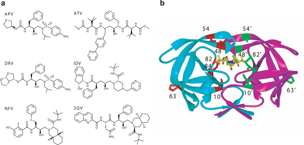
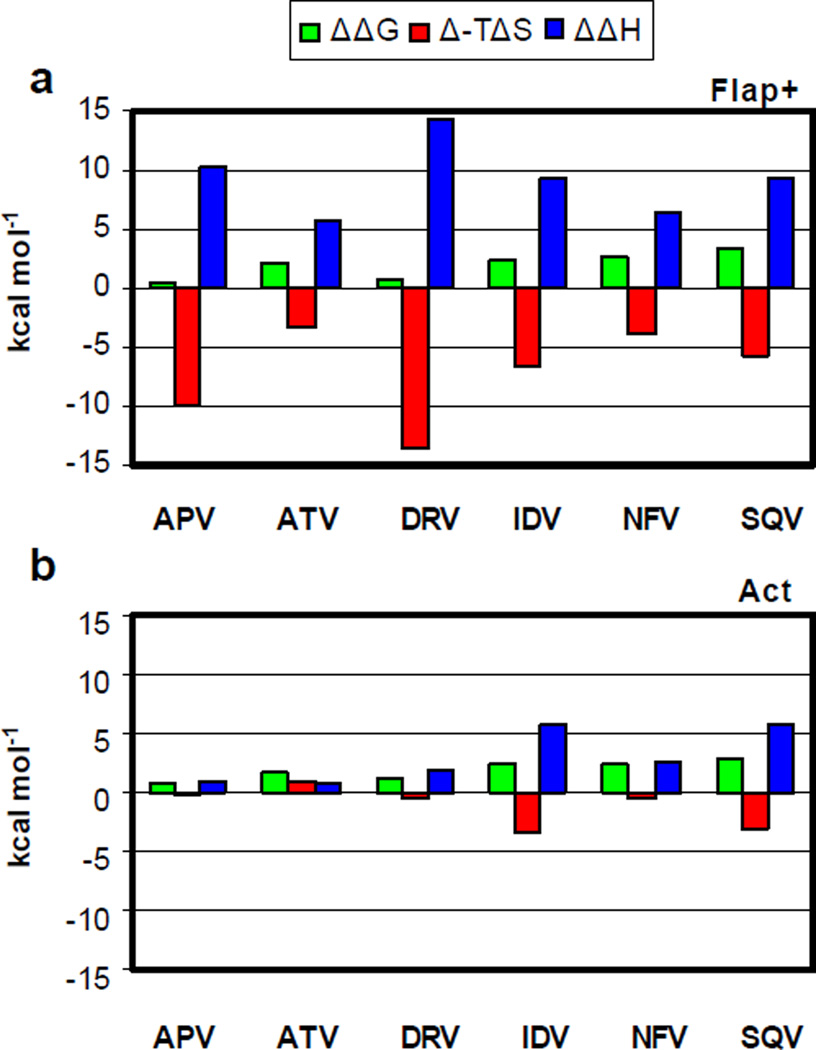
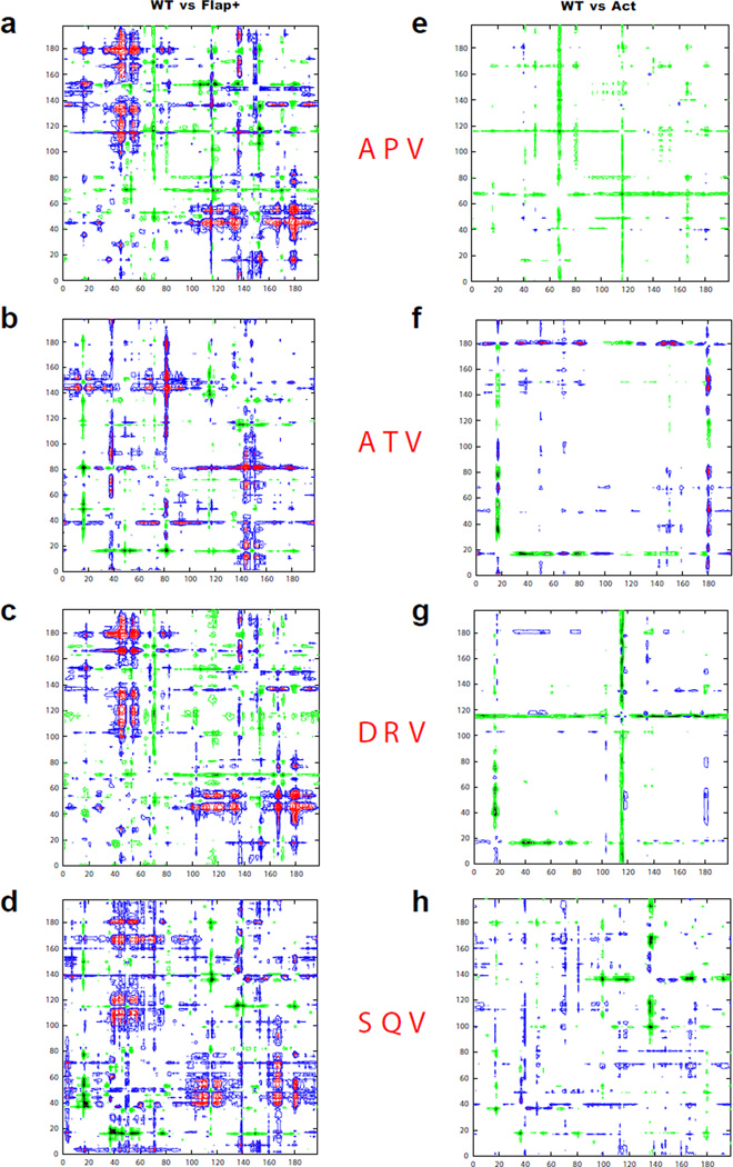
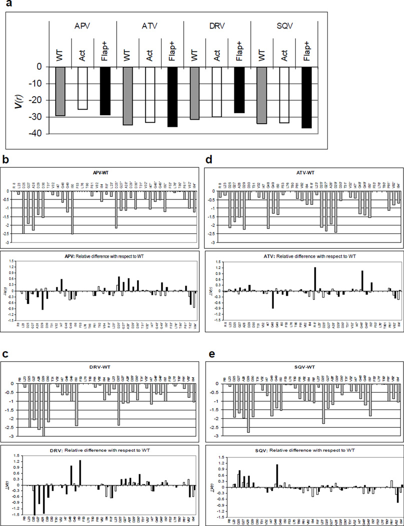
The development of HIV-1 protease inhibitors has been the historic paradigm of rational structure-based drug design, where structural and thermodynamic analyses have assisted in the discovery of novel inhibitors. While the total enthalpy and entropy change upon binding determine the affinity, often the thermodynamics are considered in terms of inhibitor properties only. In the current study, profound changes are observed in the binding thermodynamics of a drug-resistant variant compared to wild-type HIV-1 protease, irrespective of the inhibitor bound. This variant (Flap+) has a combination of flap and active site mutations and exhibits extremely large entropy-enthalpy compensation compared to wild-type protease, 5-15 kcal/mol, while losing only 1-3 kcal/mol in total binding free energy for any of six FDA-approved inhibitors. Although entropy-enthalpy compensation has been previously observed for a variety of systems, never have changes of this magnitude been reported. The co-crystal structures of Flap+ protease with four of the inhibitors were determined and compared with complexes of both the wild-type protease and another drug-resistant variant that does not exhibit this energetic compensation. Structural changes conserved across the Flap+ complexes, which are more pronounced for the flaps covering the active site, likely contribute to the thermodynamic compensation. The finding that drug-resistant mutations can profoundly modulate the relative thermodynamic properties of a therapeutic target independent of the inhibitor presents a new challenge for rational drug design.
Figures

Similar articles
-
A structural and thermodynamic escape mechanism from a drug resistant mutation of the HIV-1 protease.Proteins. 2004 May 15;55(3):594-602. doi: 10.1002/prot.20069. Proteins. 2004. PMID: 15103623
-
Thermodynamic basis of resistance to HIV-1 protease inhibition: calorimetric analysis of the V82F/I84V active site resistant mutant.Biochemistry. 2000 Oct 3;39(39):11876-83. doi: 10.1021/bi001013s. Biochemistry. 2000. PMID: 11009599
-
Overcoming drug resistance in HIV-1 chemotherapy: the binding thermodynamics of Amprenavir and TMC-126 to wild-type and drug-resistant mutants of the HIV-1 protease.Protein Sci. 2002 Aug;11(8):1908-16. doi: 10.1110/ps.0206402. Protein Sci. 2002. PMID: 12142445 Free PMC article.
-
Structural and thermodynamic basis of resistance to HIV-1 protease inhibition: implications for inhibitor design.Curr Drug Targets Infect Disord. 2003 Dec;3(4):311-28. doi: 10.2174/1568005033481051. Curr Drug Targets Infect Disord. 2003. PMID: 14754432 Review.
-
Structural and Functional Studies on HIV Protease: Mechanism of Action, Subtypes, Inhibitors, and Drug Resistance.Methods Mol Biol. 2025;2867:185-200. doi: 10.1007/978-1-0716-4196-5_11. Methods Mol Biol. 2025. PMID: 39576582 Review.
Cited by
-
An insight to the molecular interactions of the FDA approved HIV PR drugs against L38L↑N↑L PR mutant.J Comput Aided Mol Des. 2018 Mar;32(3):459-471. doi: 10.1007/s10822-018-0099-9. Epub 2018 Feb 3. J Comput Aided Mol Des. 2018. PMID: 29397520
-
Differential Flap Dynamics in Wild-type and a Drug Resistant Variant of HIV-1 Protease Revealed by Molecular Dynamics and NMR Relaxation.J Chem Theory Comput. 2012 Oct 9;8(10):3452-3462. doi: 10.1021/ct300076y. Epub 2012 Apr 17. J Chem Theory Comput. 2012. PMID: 23144597 Free PMC article.
-
Strong Ligand-Protein Interactions Derived from Diffuse Ligand Interactions with Loose Binding Sites.Biomed Res Int. 2015;2015:746980. doi: 10.1155/2015/746980. Epub 2015 May 4. Biomed Res Int. 2015. PMID: 26064949 Free PMC article.
-
A combined 3D-QSAR and docking studies for the In-silico prediction of HIV-protease inhibitors.Chem Cent J. 2013 May 17;7(1):88. doi: 10.1186/1752-153X-7-88. Chem Cent J. 2013. PMID: 23683267 Free PMC article.
-
Electrostatic Interactions between CSTF2 and pre-mRNA Drive Cleavage and Polyadenylation.Biophys J. 2022 Feb 15;121(4):607-619. doi: 10.1016/j.bpj.2022.01.005. Epub 2022 Jan 26. Biophys J. 2022. PMID: 35090899 Free PMC article.
References
-
- Chaires JB. Annual Review of Biophysics. Palo Alto: Annual Reviews; 2008. Calorimetry and thermodynamics in drug design; pp. 135–151. - PubMed
-
- Wlodawer A, Erickson JW. Structure-based inhibitors of HIV-1 protease. Ann. Rev. of Biochem. 1993;62:543–585. - PubMed
-
- Todd MJ, Luque I, Velazquez-Campoy A, Freire E. Thermodynamic basis of resistance to HIV-1 protease inhibition: calorimetric analysis of the V82F/I84V active site resistant mutant. Biochemistry. 2000;39:11876–11883. - PubMed
-
- Valzaquez-Campoy A, Todd MJ, Freire E. HIV-1 protease inhibitors: enthalpic versus entropic optimization of the binding affinity. Biochemistry. 2000;39:2201–2207. - PubMed
Publication types
MeSH terms
Substances
Grants and funding
LinkOut - more resources
Full Text Sources
Other Literature Sources
