. 2012 Sep:102:28-37.
doi: 10.1016/j.exer.2012.06.004. Epub 2012 Jun 17.

The unfolded protein response is activated in connexin 50 mutant mouse lenses

Affiliations

The unfolded protein response is activated in connexin 50 mutant mouse lenses

Bhagwat V Alapure et al. Exp Eye Res. 2012 Sep.

Abstract

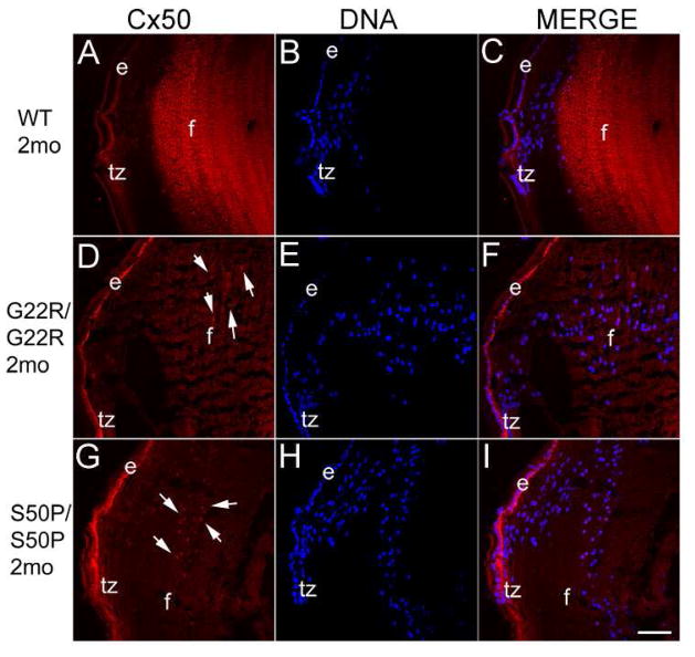
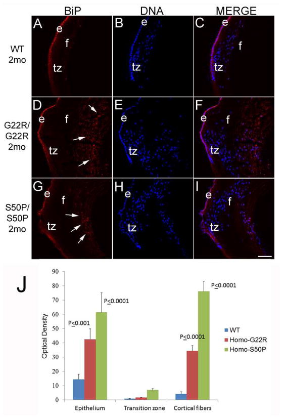
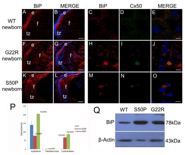
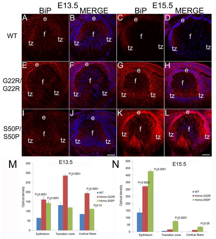
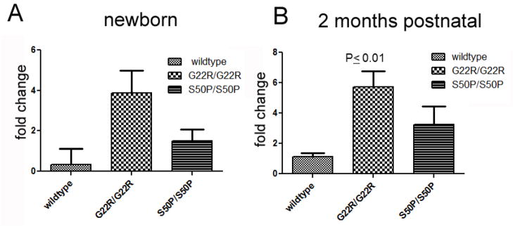
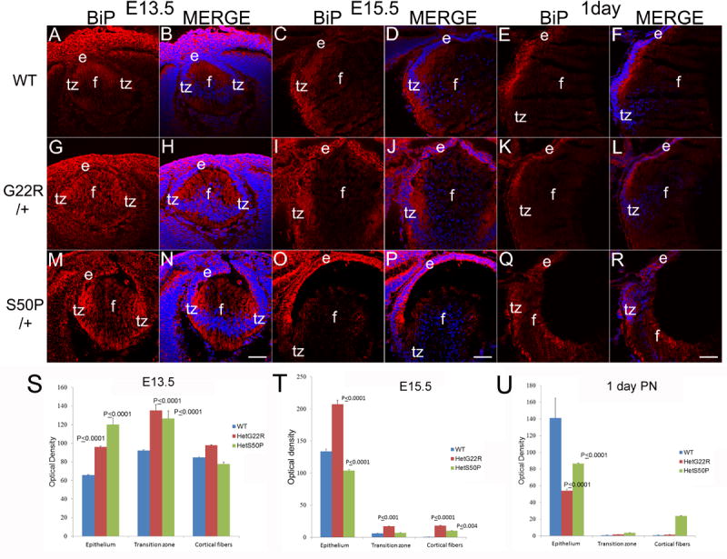
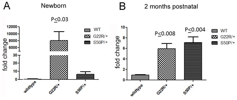
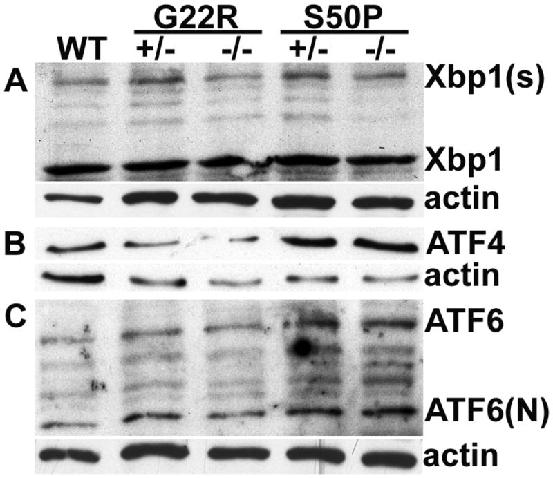
The unfolded protein response is a set of cell signaling pathways recently recognized to be activated in the lens during both normal development and endoplasmic reticulum stress induced by either unfolded proteins or oxidative damage. While mutations in the gene for connexin 50 are known to cause autosomal dominant cataracts, it has not been previously reported whether mutant connexins can activate the unfolded protein response in the lens. Mice homozygous for the S50P or G22R mutation of connexin 50 have reduced amounts of connexin 50 protein at the cell membrane, with some intracellular staining consistent with retention in the endoplasmic reticulum. Connexin 50 mutants have elevated levels of BiP expression in both lens epithelial and fiber cells from E15.5 with the most robust elevation detected in newborns. While this elevation decreases in magnitude postnatally, BiP expression is still abnormally high in adults, particularly in the perinuclear endoplasmic reticulum of cell nuclei that are inappropriately retained in adult homozygous mutant lenses. Xbp1 splicing was elevated in lenses from both connexin mutants studied, while Atf4 and Atf6 levels were not majorly affected. Overall, these data suggest that UPR may be a contributing factor to the phenotype of connexin 50 mutant lenses even though the relatively modest extent of the response suggests that it is unlikely to be a major driver of the pathology.


Figures

Figure 1
Figure 1
Darkfield imaging demonstrates that Cx50 mutant lenses have profound opacities and are smaller than normal at two months of age. A) wild type C57Bl/6 lens B) homozygous Cx50 G22R lens C) homozygous Cx50 S50P lens. Scale bar = 1 mm
Figure 2
Figure 2
Immunostaining of two month old mouse lenses with an antibody raised against the C-terminus of Cx50. Cx50 is strongly localized to the lens fiber cell membranes in WT lenses (A) while Cx50 protein is less sharply localized in homozygous Cx50 G22R (D) and S50P (G) mutants. Further, homozygous Cx50G22R (D) and Cx50S50P (G) lenses also exhibit appreciable levels of Cx50 protein surrounding the nuclear envelope (arrows). Red-Cx50; Blue-DRAQ5 (DNA); e-epithelial cells; f- fiber cells; tz-transition zone; Scale bar=77μm
Figure 3
Figure 3
BiP, a molecular chaperone found in the ER, is more abundant around the cell nuclei of maturing lens fibers in two month old Cx50 mutant mice (arrows). In wildtype lenses (A, B, C), BiP is predominately seen in the lens epithelium, with lower levels distributed through the lens fibers. In contrast, BiP staining is more prominent in maturing lens fibers, particularly in the perinuclear ER (arrows) in both Cx50 G22R (D, E, F,) and Cx50 S50P (G, H,I) homozygous mutant lenses. J- Quantitation of BiP levels in the lens epithelium, transition zone and cortical fibers using ImageJ. P values reported represent ANOVA analysis performed on results obtained from at least three biological replicates. Red-BiP; Blue-DRAQ5 (DNA); e-epithelial cells; f- fiber cells; tz-transition zone; Scale bar=77μm
Figure 4
Figure 4
BiP levels are elevated in newborn Cx50 mutant lenses (A, B,) Wildtype newborn lenses express BiP prominently in the lens epithelium with lower amounts in the lens fibers that have not completed organelle degradation. (C,D,E)- BiP and Cx50 are not found co-localized in wildtype lens fibers at birth (F, G,) Homozygous Cx50G22R lenses have increased levels of BiP expression, particularly in the lens fiber cells. (H,I,J)- The elevated amounts of BiP found in G22R lens fibers is co-localized with Cx50 (K,L) Cx50S50P lenses also have increased levels of BiP in the lens fibers although this increase is more confined to the cortical fiber cells and this additional BiP co-localizes with Cx50 (M,N,O). (P) Quantitation of BiP levels in G22R/G22R and S50P/S50P homozygotes in the lens epithelium, transition zone and cortical fibers using ImageJ. P values reported represent ANOVA analysis performed on results obtained from at least three biological replicates. (Q) Western blotting analysis of newborn lenses confirming the increase in BiP expression in the Cx50S50P and Cx50G22R mutants. Red-BiP; Blue-DRAQ5(DNA); Green- Cx50 e- epithelial cells; f- fiber cells; tz-transition zone; Scale bar A,B,F,G,K,L=77 μm, C,D,E,H,I,J,M,N,O= 6 μm
Figure 5
Figure 5
BiP upregulation begins in Cx50 mutant lenses between E13.5 and E15.5. (A, B) WT E13.5 lenses exhibit a uniform BiP distribution which is also seen in E13.5 G22R/G22R (E,F) and E13.5 S50P/S50P (I, J) connexin mutants. In WT lenses, BiP levels begin to be relatively higher in the transition zone of E15.5 lenses (C, D), BiP levels are noticeably upregulated by E15.5 in both G22R/G22R (G, H) and S50P/S50P (K,L) lenses. (M,N) Quantitation of BiP levels in G22R/G22R and S50P/S50P homozygotes in the lens epithelium, transition zone and cortical fibers at E13.5 (M) and E15.5 (N) using ImageJ. P values reported represent ANOVA analysis performed on results obtained from at least three biological replicates. Red-BiP; Blue-DRAQ5 (DNA); e- epithelial cells; f- fiber cells; tz-transition zone; Panels A,B,E,F,I,J Scale bar 67 μm; Panels C,D,G,H,K,L Scale bar=71 μm.
Figure 6
Figure 6
Quantitative rt-PCR analysis of Xbp-1 splicing in homozygous G22R and S50P mutant lenses at birth and two months postnatal. P values are obtained from unpaired T-test.
Figure 7
Figure 7
BiP levels are upregulated prenatally in heterozygous connexin mutant lenses (A,B) WT E13.5 lenses exhibit a uniform BiP distribution which is more intense in E13.5 G22R/+ (G,H) and E13.5 S50P/+ (M, N) connexin mutants. In WT lenses, BiP levels begin to be relatively higher in the transition zone of E15.5 lenses (C, D), which is similar to that seen in both G22R/+ (I, J) and S50P/+ (O,P) lenses although BiP levels may be elevated in the lens epithelium of both heterozygous mutants. By birth, BiP levels/distribution is similar between WT (E,F), G22R/+ (K,L) and S50P/+ lenses (Q,R). (S,T,U) Quantitation of BiP levels in G22R and S50P heterozygotes in the lens epithelium, transition zone and cortical fibers at E13.5 (S), E15.5 (T) and birth (U) using ImageJ. P values reported represent ANOVA analysis performed on results obtained from at least three biological replicates. Red-BiP; Blue-DRAQ5(DNA); e- epithelial cells; f- fiber cells; tz-transition zone; Scale bar 77 μm
Figure 8
Figure 8
Quantitative rt-PCR analysis of Xbp-1 splicing in heterozygous G22R and S50P mutant lenses. P values are obtained from unpaired T-test.
Figure 9
Figure 9
Western blotting analysis of XBP1, ATF4 and ATF6 in newborn heterozygous (+/−) and homozygous (−/−) Cx50S50P and Cx50G22R mutant lens. (A) Levels of protein derived from the spliced XBP1 message (Xbp1(s)) are apparently increased in the both heterozygous mutants although western blotting did not detect consistent differences in levels in homozygous mutants. (B) ATF4 levels are moderately higher in Cx50 S50P mutants and while no changes were seen for the Cx50 G22R mutant as compared to wildtype. (C) Atf6 protein levels are slightly higher in Cx50 S50P mutant lenses while no consistent changes were detected for Cx50 G22R mutants as compared to wildtype.

References

    1. Aragon T, van Anken E, Pincus D, Serafimova IM, Korennykh AV, Rubio CA, Walter P. Messenger RNA targeting to endoplasmic reticulum stress signalling sites. Nature. 2009;457:736–740. - PMC - PubMed
    1. Arora A, Minogue PJ, Liu X, Addison PK, Russel-Eggitt I, Webster AR, Hunt DM, Ebihara L, Beyer EC, Berthoud VM, Moore AT. A novel connexin50 mutation associated with congenital nuclear pulverulent cataracts. J Med Genet. 2008;45:155–160. - PMC - PubMed
    1. Austin RC. The unfolded protein response in health and disease. Antioxid Redox Signal. 2009;11:2279–2287. - PubMed
    1. Bassnett S. The fate of the Golgi apparatus and the endoplasmic reticulum during lens fiber cell differentiation. Invest Opthalmol Vis Sci. 1995;36:1793–1803. - PubMed
    1. Bassnett S, Wilmarth PA, David LL. The membrane proteome of the mouse lens fiber cell. Mol Vis. 2009;15:2448–2463. - PMC - PubMed

Publication types

MeSH terms