. 2012 Jul 1;26(13):1433-44.
doi: 10.1101/gad.193904.112. Epub 2012 Jun 19.

MUT-16 promotes formation of perinuclear mutator foci required for RNA silencing in the C. elegans germline

Affiliations

MUT-16 promotes formation of perinuclear mutator foci required for RNA silencing in the C. elegans germline

Carolyn M Phillips et al. Genes Dev. .

Abstract

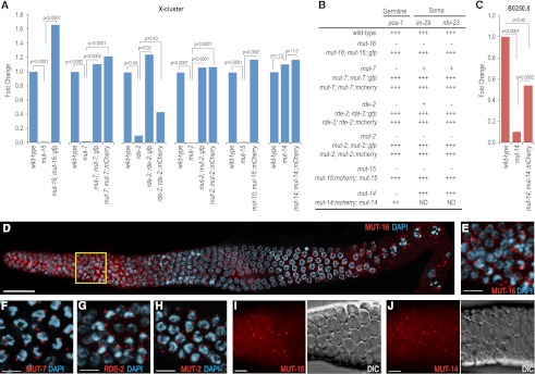
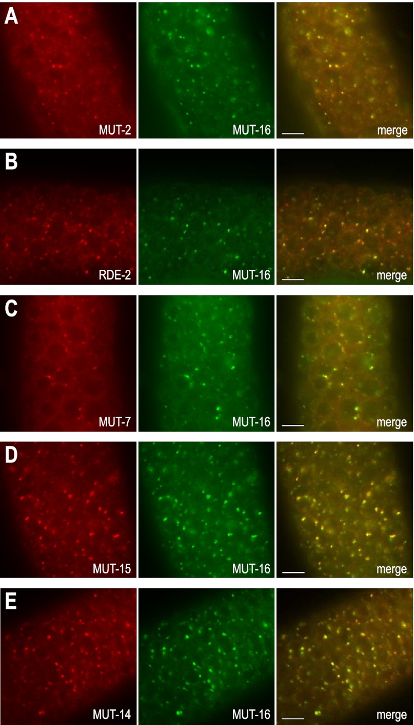
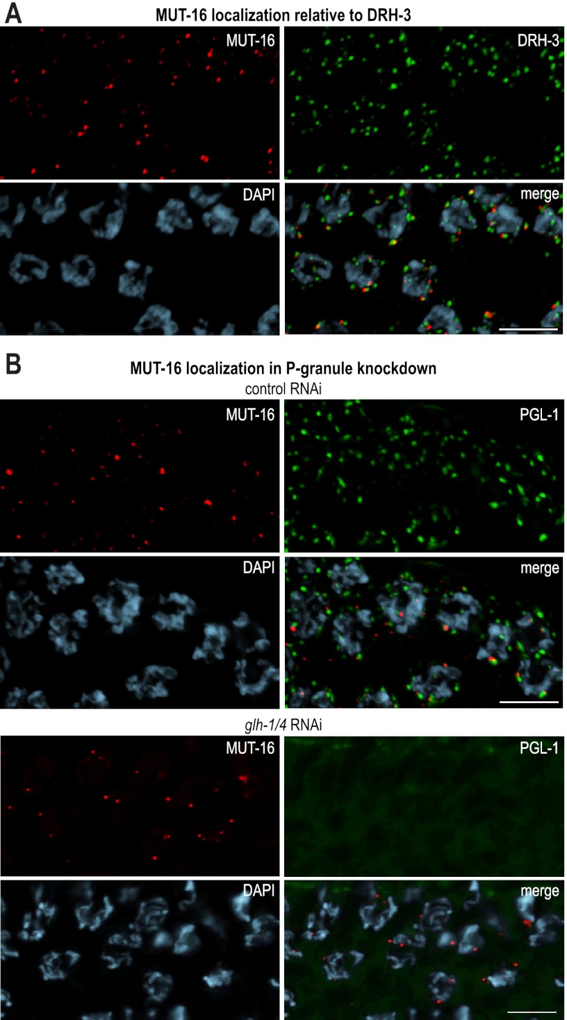
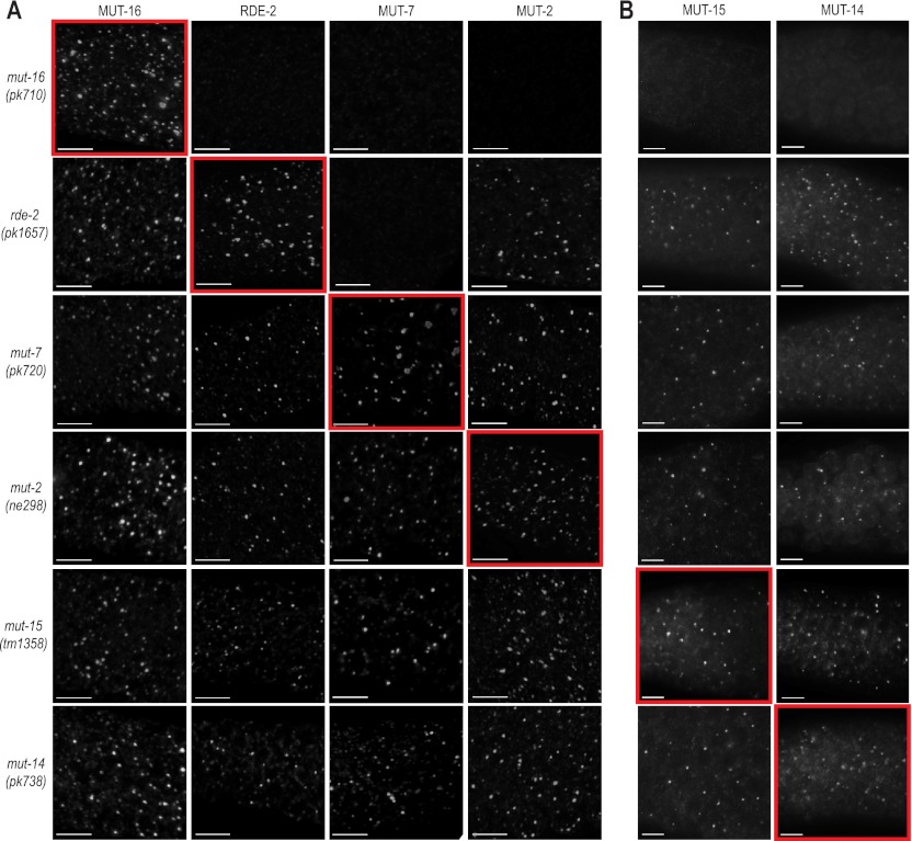
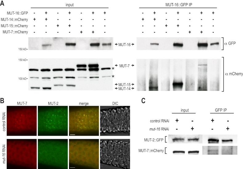
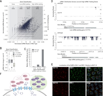
RNA silencing can be initiated by endogenous or exogenously delivered siRNAs. In Caenorhabditis elegans, RNA silencing guided by primary siRNAs is inefficient and therefore requires an siRNA amplification step involving RNA-dependent RNA polymerases (RdRPs). Many factors involved in RNA silencing localize to protein- and RNA-rich nuclear pore-associated P granules in the germline, where they are thought to surveil mRNAs as they exit the nucleus. Mutator class genes are required for siRNA-mediated RNA silencing in both germline and somatic cells, but their specific roles and relationship to other siRNA factors are unclear. Here we show that each of the six mutator proteins localizes to punctate foci at the periphery of germline nuclei. The Mutator foci are adjacent to P granules but are not dependent on core P-granule components or other RNAi pathway factors for their formation or stability. The glutamine/asparagine (Q/N)-rich protein MUT-16 is specifically required for the formation of a protein complex containing the mutator proteins, and in its absence, Mutator foci fail to form at the nuclear periphery. The RdRP RRF-1 colocalizes with MUT-16 at Mutator foci, suggesting a role for Mutator foci in siRNA amplification. Furthermore, we demonstrate that genes that yield high levels of siRNAs, indicative of multiple rounds of siRNA amplification, are disproportionally affected in mut-16 mutants compared with genes that yield low levels of siRNAs. We propose that the mutator proteins and RRF-1 constitute an RNA processing compartment required for siRNA amplification and RNA silencing.


Figures

Figure 1.
Figure 1.
Mutator proteins localize to perinuclear foci in the germline. (A) Fluorescently tagged mutator proteins rescue mutations in their respective genes as assayed by TaqMan quantitative RT–PCR to detect the X-Cluster siRNA 22G siR-1. The mean is calculated from two biological replicates for each strain. (B) Susceptibility of mutator mutant worms to germline and somatic RNAi in the presence and absence of rescuing transgenes. pos-1 RNAi scored as complete embryonic lethality (that is, normal response to pos-1 RNAi) (+++), complete embryonic viability (that is, an RNAi-defective response) (−), or ∼50% embryonic viability (++). lin-29 RNAi scored as 100% vulval bursting (+++), 100% viable adults (−), or adults with morphological defects (i.e., protruding vulva) (+). nhr-23 RNAi scored as 100% larval arrest (+++), 100% viable adults (−), or adults with morphological defects (+). (C) mCherry-tagged MUT-14 rescues the mut-14(pk738) mutant as assayed by TaqMan quantitative RT–PCR of the germline-expressed B0250.8 siRNA. The mean is calculated from two biological replicates for each strain. (D) MUT-16 (red) localizes throughout the germline, but is brightest in the mitotic proliferation and transition zone regions as well as in the diplotene/diakinesis stages of meiosis. The yellow box is magnified in E. Image depicts entire dissected gonad stained with DAPI (blue) and anti-GFP (recognizing MUT-16::GFP). Image is an assembly of four three-dimensional (3D) data stacks following deconvolution. Bars, 20 μm. (E–J) Mutator proteins localize to foci in the germlines of adult hermaphrodites. MUT-16::GFP (E), MUT-7::GFP (F), RDE-2::GFP (G), and MUT-2::GFP (H) associate with the nuclear periphery as visualized by DAPI staining (blue). MUT-15::mCherry (I) and MUT-14::mCherry (J) mCherry fluorescence images are displayed next to the corresponding DIC images. All animals were dissected prior to imaging. Bars, 5 μm.
Figure 2.
Figure 2.
MUT-16 colocalizes with other mutator proteins at germline foci. MUT-2::mCherry (A), RDE-2::mCherry (B), MUT-7::mCherry (C), MUT-15::mCherry (D), and MUT-14::mCherry (E) colocalize with MUT-16::GFP. All animals were dissected prior to imaging. Bars, 5 μm.
Figure 3.
Figure 3.
Mutator proteins localize independently of P-granule components. (A) MUT-16 (red) and DRH-3 (green) form distinct foci adjacent to germline nuclei. Staining was performed using antibodies against GFP (MUT-16 in red), DRH-3 (green), and DAPI (blue). (B) MUT-16 and PGL-1 foci partially overlap in adult C. elegans feeding on Escherichia coli expressing control (empty vector) dsRNA. Upon treatment with glh-1/glh-4 dsRNA, PGL-1 becomes diffuse, but MUT-16 is unchanged. Proteins were visualized using anti-GFP (MUT-16 in red) and anti-dsRed (PGL-1 in green). DNA was stained by DAPI (blue). Bars, 5 μm.
Figure 4.
Figure 4.
Genetic requirements for mutator protein localization. (A) MUT-16::GFP, RDE-2::GFP, MUT-7::GFP, and MUT-2::GFP expression in each of the mutator mutants. Images highlighted by red boxes display expression from a transgene in the corresponding mutant. All animals were dissected and stained with anti-GFP. Bars, 5 μm. (B) MUT-15::mCherry or MUT-14::mCherry were introduced into each of the six mutator mutants. Images highlighted by red boxes display expression from a transgene in the corresponding mutant. Bars, 5 μm.
Figure 5.
Figure 5.
MUT-16 is essential for mutator complex formation. (A) GFP and mCherry proteins from total lysate (input, left panels) and GFP-IP (right panels) from the indicated transgenic strains as assayed by Western blot. MUT-14::mCherry (93.1 kDa), MUT-15::mCherry (96.1 kDa), and MUT-7::mCherry (139.5 kDa) coimmunoprecipitate with MUT-16::GFP (154.4 kDa). Asterisks mark bands resulting from cross-reactivity of anti-mCherry with non-mutator proteins. (B) MUT-7::mCherry (red) and MUT-2::GFP (green) expression in C. elegans treated with control or mut-16 RNAi. (C) GFP and mCherry proteins from total lysate (input, left panels) and GFP-IP (right panels) from the transgenic strain containing MUT-2::GFP (86.6 kDa) and MUT-7::mCherry (139.5 kDa) treated with control or mut-16 RNAi and assayed by Western blot.
Figure 6.
Figure 6.
MUT-16 is required for siRNA amplification. (A) Scatter plots display small RNA RPM on a log2 scale for each annotated coding gene in wild-type (bottom axis) and mut-16 mutants (left axis). The fold reduction of siRNA reads in mut-16 mutants relative to wild type is indicated by the diagonal lines on the right axis. (B) Box plots display ratio of siRNA reads on a log2 scale in mut-16 relative to wild type for low siRNA yielding genes (1–10 RPM or 10–100 RPM) and high siRNA yielding genes (>100 RPM). (C) Median siRNA reads per gene for low siRNA yielding genes (1–10 RPM or 10–100 RPM) or high siRNA yielding genes (>100 RPM) in wild-type and mut-16 mutants. (D) Small RNA distribution across the low siRNA yielding gene aagr-3 and the high siRNA yielding gene B0250.8 in wild-type and mut-16 mutants. (E) Localization of HA::EGO-1 and Flag::RRF-1 relative to MUT-16::GFP in dissected germlines immunostained with anti-GFP and either anti-HA or anti-Flag antibodies. Bars, 5 μm. (F) Model depicting the composition and localization of Mutator foci and P granules adjacent to nuclear pores.

References

    1. Aravin AA, van der Heijden GW, Castañeda J, Vagin VV, Hannon GJ, Bortvin A 2009. Cytoplasmic compartmentalization of the fetal piRNA pathway in mice. PLoS Genet 5: e1000764 doi: 10.1371/journal.pgen.1000764 - PMC - PubMed
    1. Batista PJ, Ruby JG, Claycomb JM, Chiang R, Fahlgren N, Kasschau KD, Chaves DA, Gu W, Vasale JJ, Duan S, et al. 2008. PRG-1 and 21U-RNAs interact to form the piRNA complex required for fertility in C. elegans. Mol Cell 31: 67–78 - PMC - PubMed
    1. Bernstein E, Caudy AA, Hammond SM, Hannon GJ 2001. Role for a bidentate ribonuclease in the initiation step of RNA interference. Nature 409: 363–366 - PubMed
    1. Brenner S 1974. The genetics of Caenorhabditis elegans. Genetics 77: 71–94 - PMC - PubMed
    1. Chen C-CG, Simard MJ, Tabara H, Brownell DR, McCollough JA, Mello CC 2005. A member of the polymerase β nucleotidyltransferase superfamily is required for RNA interference in C. elegans. Curr Biol 15: 378–383 - PubMed

Publication types

MeSH terms

Substances