. 2012 Jul 10;107(2):315-24.
doi: 10.1038/bjc.2012.263. Epub 2012 Jun 19.

Mixed lineage leukaemia-4 regulates cell-cycle progression and cell viability and its depletion suppresses growth of xenografted tumour in vivo

Affiliations

Mixed lineage leukaemia-4 regulates cell-cycle progression and cell viability and its depletion suppresses growth of xenografted tumour in vivo

K I Ansari et al. Br J Cancer. .

Abstract

Background: Mixed lineage leukaemia-4 (MLL4) is one of the MLL family of histone H3 lysine-4 (H3K4)-specific methyl transferases that have critical roles in gene expression and epigenetics in human. Though MLLs are well recognised as crucial players in histone methylation and gene regulation; little is known about the biochemical functions of MLL4 and its roles in cancer.

Methods: Herein, we have investigated the roles of MLL4 in cell viability, cell-cycle progression and explored its potential roles in tumour growth using antisense-mediated knockdown experiments, flow-cytometry analysis, chromatin immunoprecipitation, immunofluorescence staining and animal models.

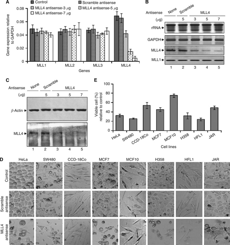
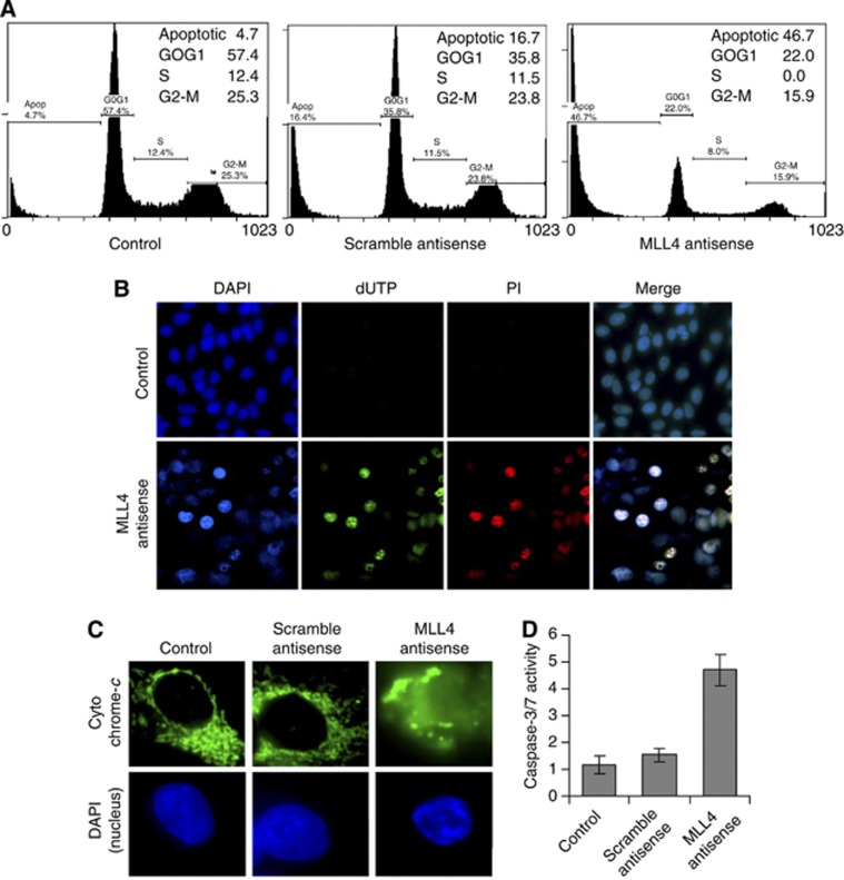
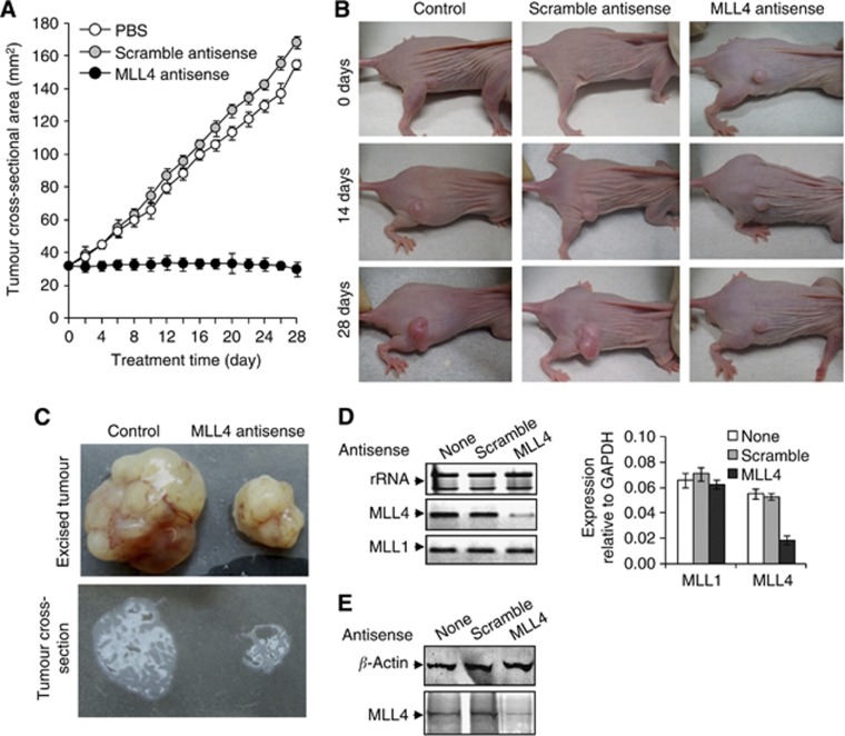
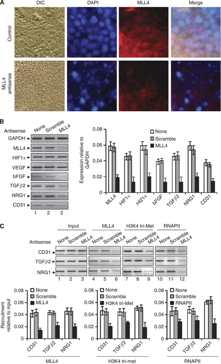
Results: Our studies demonstrated that knockdown of MLL4 severely affects cell-cycle progression and induces apoptotic cell death in cultured tumour cells. Knockdown of MLL4 induced nuclear condensation, fragmentation, cytochrome-c release from mitochondria to cytosol and activated caspase-3/7 indicating apoptotic cell death. The MLL4 regulates expression of various critical cell-cycle regulatory genes such as cyclin D, cyclin E, p27, HOXA5 and HOXB7 via histone H3K4 trimethylation and recruitment of RNA polymerase II. Interestingly, application of MLL4 antisense suppressed tumour growth in vivo in colon cancer xenograft implanted in nude mouse. The MLL4 antisense specifically knocked down MLL4 in tumour tissue and also downregulated the expression of various growth and angiogenic factors resulting in tumour suppression.

Conclusion: Our results demonstrated that MLL4 is a crucial player in cell viability, cell-cycle progression and is critical for tumour growth in vivo.


Figures

Figure 1
Figure 1
MLL4 knockdown and its effect on cell viability. (A) Effect of MLL4 knockdown on the expression of MLL1–4: colon cancer (SW480) cells were transfected with varying concentrations of MLL4 antisense for 48 h. RNA was reverse transcribed and subjected to real-time PCR analysis using primers specific to MLL1, MLL2, MLL3, MLL4 and GAPDH. The expression of different MLLs relative to GAPDH was plotted. GAPDH expression and rRNA levels were used as loading control. Each experiment was performed in three replicates for at least two times. Bars indicate standard errors. (B) RNA from the control and MLL4 antisense-treated cells were subjected to RT–PCR using primer specific to MLL4, MLL1 (specificity control) and GAPDH (loading control) and then analysed by agarose gel electrophoresis. Lane 1: control cells; lane 2: cells transfected with scramble antisense; lanes 3–5: cell transfected with 3–7 μg of MLL4 antisense. (C) Western blotting: proteins from the control and MLL4 antisense-treated cells were subjected to western blotting using anti-MLL4 and anti-β-actin antibodies (loading control). (D) Microscopic images showing different cancer and non-cancer cells transfected with MLL4 or scramble antisense for 48 h. Control cells were treated with PBS (buffer) alone. (E) MTT assay: cells were transfected with 7 μg MLL4 or scramble antisense for 48 h and then subjected to MTT assay. The relative (%) cell viability (MLL4 antisense vs scramble antisense) was plotted for different cell lines. Bars indicate standard errors.
Figure 2
Figure 2
MLL4 knockdown induced apoptosis in colon cancer cell. SW480 cells were transfected with MLL4 antisense or scramble for 48 h then subjected to different analysis. (A) FACS analysis: MLL4 and scramble antisense-transfected cell was fixed, stained with propidium iodide (PI) and analysed by using Beckman Coulter Cytomics FC500 flow cytometry analyser. Percent cell populations at different stages of cell cycle are listed within the panels. (B) TUNEL assay. MLL4 antisense-treated and control cells were subjected to terminal nicked end-labelling using fluorescent dUTP, stained with DAPI (nuclear staining, blue fluorescence) and PI (PI that stains nucleus of dead cells, red colour) and analysed under fluorescence microscope. dUTP-stained green speckles represent apoptotic cells with fragmented nuclei. (C) Immunostaining of cytochrome-c: MLL4 antisense-treated and control cells were immunostained with cytochrome-c antibody followed by FITC-labelled secondary antibody, stained with DAPI and visualised under fluorescence microscope. (D) Caspase analysis: MLL4 antisense (or scramble antisense) cell lysates were analysed for caspase activity using Caspase-3/7 assay kit. Bars indicated standard error.
Figure 3
Figure 3
MLL4 regulates cell-cycle regulatory cyclins, p-protein and HOX genes. SW480 cells were transfected with MLL4 antisense for 48 h and the subjected to RNA extraction or ChIP assay. (A) RNA from MLL4-knocked down or control cells analysed by RT–PCR by using primers specific to selected cell-cycle regulatory genes (cyclin A–E, p27, HOXA5 and HOXB7) and GAPDH. GAPDH expression and rRNA levels were used as loading control. (B) ChIP assay: MLL4 and scramble antisense-treated and control cells were subjected to ChIP with MLL4, H3K4 tri-methyl, RNAPII and β-actin (control) antibodies. The immunoprecipitated DNA fragments were PCR amplified by using primers specific to the β-actin (ORF, as control) and promoters of cyclin B, cyclin D, p27, HOXA5 and HOXB7 genes. The real-time quantification is on the bottom panel. Bars indicate standard errors (n=3).
Figure 4
Figure 4
Regression of colon cancer xenograft by MLL4 antisense. SW480 cells were subcutaneously injected on the right hinge region of 6-week-old athymic nude (nu/nu) mice. Once the tumour reached about 32 mm2 of cross-sectional area, mice were intraperitoneally administered with MLL4 antisense (300 μg per 20 g body weight) on the left hinge region at 4 days interval for 4 weeks. Control mice were administered with either PBS (diluents) or a same doze of scramble antisense (with no homology with MLL4). Tumour sizes were measured using a slid caliper at every 2 days intervals. (A) Cross-sectional area of the tumours was plotted against time. Bars indicated standard errors. (B) Representative pictures of the control and antisense-treated mice at different stages. (C) Tumour xenografts excised at 28th day of treatment. The cross-section of tumours showing internal issue is in bottom panel. (D) The RNA extract of the excised tumour was analysed by RT–PCR with primer specific to MLL4 and MLL1 (specificity control). rRNA was used as quantitative control. The real-time quantification is on the right panel. Bars indicate standard errors (n=3). (E) The western blot of the protein extract of the excised tumour was done with MLL4 and β-actin (control) antibody.
Figure 5
Figure 5
MLL4 knockdown affects nuclear integrity and expression of growth and angiogenic factors in xenografted tumour. The MLL4 antisense-treated or control mice with colon cancer xenograft were either euthanised to excise the tumour xenograft or perfused with 4% paraformaldehyde at 28th day of treatment. (A) Immunolocalisation of MLL4 and nuclear integrity: Formaldehyde-fixed tumours were sectioned, immunostained with MLL4 antibody and DAPI (for nuclear staining) and analysed under fluorescence microscope. Representative images showing the cellular morphology (DIC), nuclear integrity (DAPI) and MLL4 expression in the control and MLL4 antisense-treated tumours are shown. (B) MLL4 regulates expression of growth and angiogenic factors in vivo: RT–PCR analysis of the RNA extracted from MLL4 antisense-treated tumour tissues by using primers specific to MLL4, VEGF, bFGF, TGFβ2, CD31, HIF1α and β-actin (control). The real-time quantification (relative to GAPDH) is shown in right panel. Bars indicate standard error. (C) The tumour tissue was analysed by ChIP assay using MLL4, H3K4-trimethyl and RNAP II antibodies. The ChIP DNA was analysed by PCR with primers specific to the promoter regions of CD31, TGFβ2 and NRG1. qPCR data are the right panel.

References

    1. Ajiro K, Allis CD (2002) Histone code hypothesis. Tanpakushitsu Kakusan Koso 47: 753–760 - PubMed
    1. Ansari KI, Grant JD, Woldemariam GA, Kasiri S, Mandal SS (2009) Iron(III)-salen complexes with less DNA cleavage activity exhibit more efficient apoptosis in MCF7 cells. Org Biomol Chem 7: 926–932 - PubMed
    1. Ansari KI, Hussain I, Kasiri S, Mandal SS (2012) HOXC10 is overexpressed in breast cancer and transcriptionally regulated by estrogen via involvement of histone methylases MLL3 and MLL4. J Mol Endocrinol 48: 61–75 - PubMed
    1. Ansari KI, Hussain I, Shrestha B, Kasiri S, Mandal SS (2011) HOXC6 Is transcriptionally regulated via coordination of MLL histone methylase and estrogen receptor in an estrogen environment. J Mol Biol 411: 334–349 - PMC - PubMed
    1. Ansari KI, Mandal SS (2010) Mixed lineage leukemia: roles in gene expression, hormone signaling and mRNA processing. FEBS J 277: 1790–1804 - PubMed

Publication types

MeSH terms