Review
. 2012 Sep;44(9):1519-30.
doi: 10.1016/j.biocel.2012.06.013. Epub 2012 Jun 17.

Neuromyelitis optica: aquaporin-4 based pathogenesis mechanisms and new therapies

Affiliations
Review

Neuromyelitis optica: aquaporin-4 based pathogenesis mechanisms and new therapies

Julien Ratelade et al. Int J Biochem Cell Biol. 2012 Sep.

Abstract

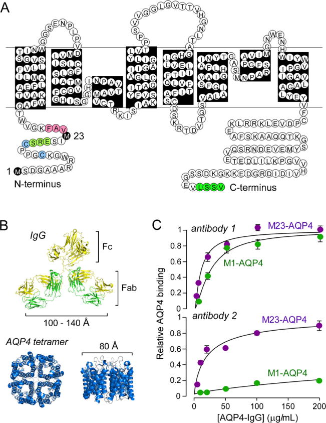
Neuromyelitis optica (NMO) is an autoimmune 'aquaporinopathy' of the central nervous system that causes inflammatory demyelinating lesions primarily in spinal cord and optic nerve, leading to paralysis and blindness. NMO lesions show loss of aquaporin-4 (AQP4), GFAP and myelin, infiltration of granulocytes and macrophages, and perivascular deposition of activated complement. Most patients with NMO are seropositive for immunoglobulin autoantibodies (AQP4-IgG) against AQP4, the principal water channel of astrocytes. There is strong evidence that AQP4-IgG is pathogenic in NMO, probably by a mechanism involving complement-dependent astrocyte cytotoxicity, causing leukocyte infiltration, cytokine release and blood-brain barrier disruption, which leads to oligodendrocyte death, myelin loss and neuron death. Here, we review the evidence for this and alternative proposed NMO pathogenesis mechanisms, such as AQP4-IgG-induced internalization of AQP4 and glutamate transporters, complement-independent cell-mediated cytotoxicity, and AQP4-IgG inhibition of AQP4 water transport function. Based on the initiating pathogenic role of AQP4-IgG binding to astrocyte AQP4 in NMO, selective blocker therapies are under development in which AQP4-targeted monoclonal antibodies or small molecules block binding of AQP4-IgG to astrocytes and consequent downstream pathology.


Figures

Fig. 1
Fig. 1
AQP4-IgG binding to its target, AQP4. (A) Amino acid sequence of human AQP4 showing Met-1 and Met-23 translation inhibition sites (black). Pink – residues involved in intermolecular N-terminus associations to form OAPs; light green – residues preventing OAP formation by M1-AQP4; blue – cysteine residues involved in palmitoylation-regulated OAP assembly; and dark green – C-terminus PDZ domain. (B) Crystal structure of human AQP4 (PDB, 3GD8) showing tetrameric association with central aqueous pore, along with structure of a generic IgG antibody shown on the same size scale. (C) Binding of two different monoclonal recombinant NMO autoantibodies to cells expressing M1-AQP4 or M23-AQP4. Parts of this figure were adapted from work originally published in the Journal of Biological Chemistry (Crane et al., 2011a) © the American Society for Biochemistry and Molecular Biology.
Fig. 2
Fig. 2
Complement-dependent cytotoxicity requires AQP4 assembly in orthogonal arrays. (A) (left) Confocal and TIRFM of M1- and M23-AQP4 transfected CHO cells immunostained for AQP4 using a C-terminus anti-AQP4 antibody. (right) SDS-PAGE (top) and BN-PAGE (bottom) of cell homogenates. (B) Cells expressing M1-AQP4 are resistant to CDC caused by AQP4-IgG in human NMO sera. (left) Cytotoxicity plotted as a function of total IgG concentration (from NMO patient serum) in the presence of human complement. Cells were incubated for 1 h prior to LDH release assay. (right) Fluorescence micrographs showing live/dead (green/red) cell staining. (C) Multivalent binding of C1q to Fc regions of clustered AQP4-IgG bound to OAP-assembled AQP4. Side-view (left) and en-face view (right) shown. Parts of this figure were adapted from work originally published in the Journal of Biological Chemistry (Phuan et al., 2012).
Fig. 3
Fig. 3. Ex vivo
organ culture models of NMO. (A) Schematic showing spinal cord slices cultured on a semi-porous membrane at an air-medium interface. After 7 days in culture, spinal cord slices were incubated with human complement (HC) and/or AQP4-IgG for 2–3 days. (B) Immunofluorescence for GFAP (green), AQP4 (red) and myelin basic protein (MBP) (red) in wildtype (AQP4+/+)and AQP4 knockout (AQP4−/−) mice.‘Control’ indicates no added AQP4-IgG or HC. (C) Schematic of optic nerve culture model showing 24 h incubation of freshly isolated optic nerves. Adapted from Zhang et al., (2011).
Fig. 4
Fig. 4
Intracerebral injection of AQP4-IgG and human complement produces NMO lesions in mice. Wildtype (AQP4+/+) and AQP4 knockout (AQP4−/−) mice were killed 4 days after injection of AQP4-IgG and human complement (HC). Brain sections were immunostained for AQP4, GFAP and myelin basic protein (MBP). Yellow lines represent needle tracts. White dashed lines demarcate lesions.
Fig. 5
Fig. 5
NMO pathogenesis mechanism. In the normal CNS, AQP4 is expressed at astrocyte end-feet facing the blood-brain barrier formed by endothelial cells connected by tight junctions (labeled ‘1’). In NMO, by an unknown mechanism, circulating AQP4-IgG crosses the blood-brain barrier and binds AQP4 on astrocytes (2). This leads to recruitment and activation of complement and deposition of the membrane attack complex (MAC), producing astrocyte damage (3). Complement activation and cytokine secretion by astrocytes recruit inflammatory cells (eosinophils, neutrophils and macrophages), which further disrupt the blood-brain barrier, allowing more entry of AQP4-IgG (4). Degranulating inflammatory cells (5) and astrocyte damage secondarily cause oligodendrocyte injury, myelin loss and axon damage (6).
Fig. 6
Fig. 6
Alternative proposed NMO pathogenesis mechanisms based on AQP4-IgG binding to AQP4. (A) Antibody-dependent cell-mediated cytotoxicity. (B) Glutamate excitotoxicity. (C) Preferential M1-AQP4 internalization. (D) AQP4 water transport inhibition. See text for explanations.
Fig. 7
Fig. 7
NMO therapy based on blocking of AQP4-IgG binding to AQP4 on astrocytes. (A) Overview of NMO pathogenesis showing AQP4-IgG production by lymphocytes, binding to AQP4 on astrocytes, CDC and (potentially) ADCC, and initiation of a cascade of inflammatory events. Therapeutic approaches shown in black boxes. (B) Schematic of AQP4-IgG antibody showing heavy (VH) and light (VL) chain variable regions, light chain constant region (CL), and heavy chain constant regions (CH1-CH3). Locations of amino acid mutations introduced in the CH2 domain of AQP4-IgG to reduce CDC (K322A), ADCC (K326W/E333S) or both (L234A/L235A). (C) Aquaporumab (AQmab) prevents CDC following exposure to AQP4-IgG and complement as shown by live/dead cell assay. Adapted with permission from Tradtrantip et al. (2012b).

References

    1. Antoine JC, Camdessanche JP, Absi L, Lassabliere F, Feasson L. Devic disease and thymoma with anti-central nervous system and antithymus antibodies. Neurology. 2004;62:978–80. - PubMed
    1. Auguste KI, Jin S, Uchida K, Yan D, Manley GT, Papadopoulos MC, et al. Greatly impaired migration of implanted aquaporin-4-deficient astroglial cells in mouse brain toward a site of injury. FASEB Journal. 2007;21:108–16. - PubMed
    1. Balbi P, Salvini S, Fundaro C, Frazzitta G, Maestri R, Mosah D, et al. The clinical spectrum of late-onset Alexander disease: a systematic literature review. Journal of Neurology. 2010;257:1955–62. - PubMed
    1. Bennett JL, Lam C, Kalluri SR, Saikali P, Bautista K, Dupree C, et al. Intrathecal pathogenic anti-aquaporin-4 antibodies in early neuromyelitis optica. Annals of Neurology. 2009;66:617–29. - PMC - PubMed
    1. Binder DK, Yao X, Zador Z, Sick TJ, Verkman AS, Manley GT. Increased seizure duration and slowed potassium kinetics in mice lacking aquaporin-4 water channels. Glia. 2006;53:631–6. - PubMed