An X-ray diffraction study on a single rod outer segment from frog retina
- PMID: 22713892
- PMCID: PMC3380658
- DOI: 10.1107/S0909049512018535
An X-ray diffraction study on a single rod outer segment from frog retina
Abstract
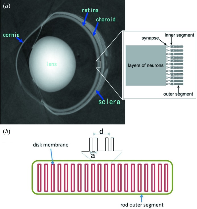
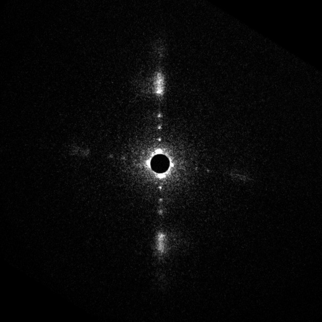
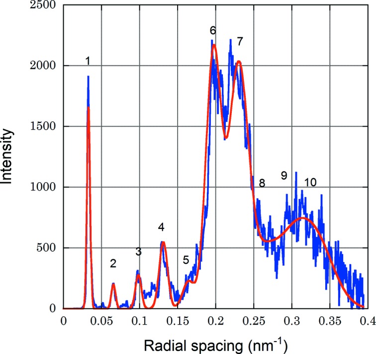
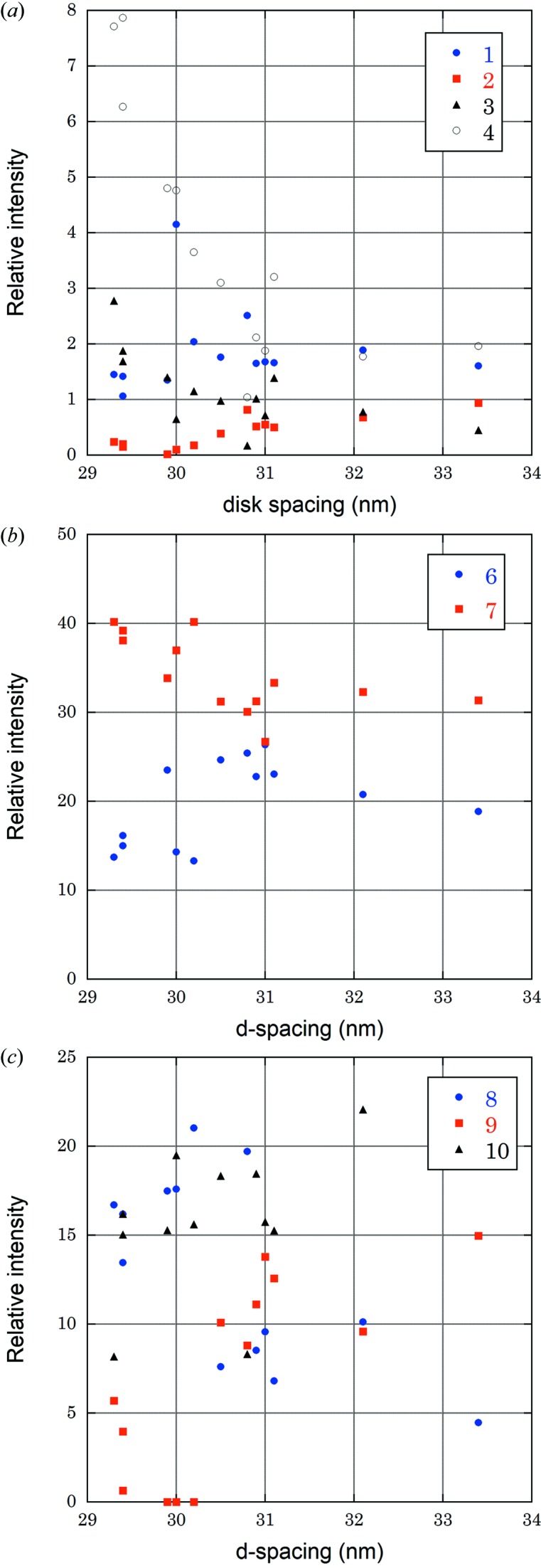
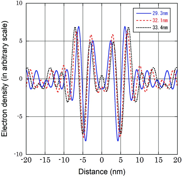
X-ray diffraction patterns were recorded from isolated single rod outer segments of frog. The outer segments in Ringer's solution were exposed to a 6 µm microbeam (15 keV) at the BL40XU beamline, SPring-8. The diffraction pattern demonstrated a remarkable regularity in the stacking and flatness of the disk membranes. The electron density profile calculated from the intensity of up to tenth-order reflections showed a pair of bilayers that comprise a disk membrane. The structure of the disk membrane and the changes in the profile on swelling generally agreed with previous reports. Radiation damage was significant with an irradiation of 5 × 10(5) Gy which is much lower than the known damaging dose on proteins at the liquid-nitrogen temperature.
Figures


Similar articles
-
A 12 A resolution X-ray diffraction study of the profile structure of isolated bovine retinal rod outer segment disk membranes.Biochim Biophys Acta. 1984 Oct 17;777(1):9-20. doi: 10.1016/0005-2736(84)90491-7. Biochim Biophys Acta. 1984. PMID: 6487620
-
[Molecular mechanisms of photoreception. V. Isolation of cytoplasmic membranes of rod outer segments from the frog retina].Mol Biol (Mosk). 1986 Nov-Dec;20(6):1479-87. Mol Biol (Mosk). 1986. PMID: 3807905 Russian.
-
Evaluation of the electron density profile of the frog rod outer segment disc-membrane in vivo using x-ray diffraction.Biochim Biophys Acta. 1981 Jan 8;640(1):142-58. doi: 10.1016/0005-2736(81)90540-x. Biochim Biophys Acta. 1981. PMID: 6971125
-
Structural changes in rod outer segments of frog and mouse after illumination.Exp Eye Res. 2013 Nov;116:395-401. doi: 10.1016/j.exer.2013.09.016. Epub 2013 Oct 3. Exp Eye Res. 2013. PMID: 24095685 Review.
-
Evidence of bilayer structure and of membrane interactions from X-ray diffraction analysis.Biochim Biophys Acta. 1982 May 12;650(4):167-207. doi: 10.1016/0304-4157(82)90016-8. Biochim Biophys Acta. 1982. PMID: 7046799 Review. No abstract available.
Cited by
-
Fiber Diffraction and Small-Angle Scattering for Structural Investigation of Bacterial Amyloids.Methods Mol Biol. 2022;2538:95-107. doi: 10.1007/978-1-0716-2529-3_7. Methods Mol Biol. 2022. PMID: 35951295
References
-
- Blasie, J. K., Worthington, C. R. & Dewey, M. M. (1969). J. Mol. Biol. 39, 407–416. - PubMed
-
- Blaurock, A. E. & Wilkins, M. H. F. (1969). Nature (London), 223, 906–909. - PubMed
-
- Blaurock, A. E. & Wilkins, M. H. F. (1972). Nature (London), 236, 313–314. - PubMed
-
- Chabre, M. (1975). Biochim. Biophys. Acta, 382, 322–335. - PubMed
MeSH terms
LinkOut - more resources
Full Text Sources