

Altered global and regional brain mean diffusivity in patients with obstructive sleep apnea
- PMID: 22715089
- PMCID: PMC3418429
- DOI: 10.1002/jnr.23083
Altered global and regional brain mean diffusivity in patients with obstructive sleep apnea
Abstract
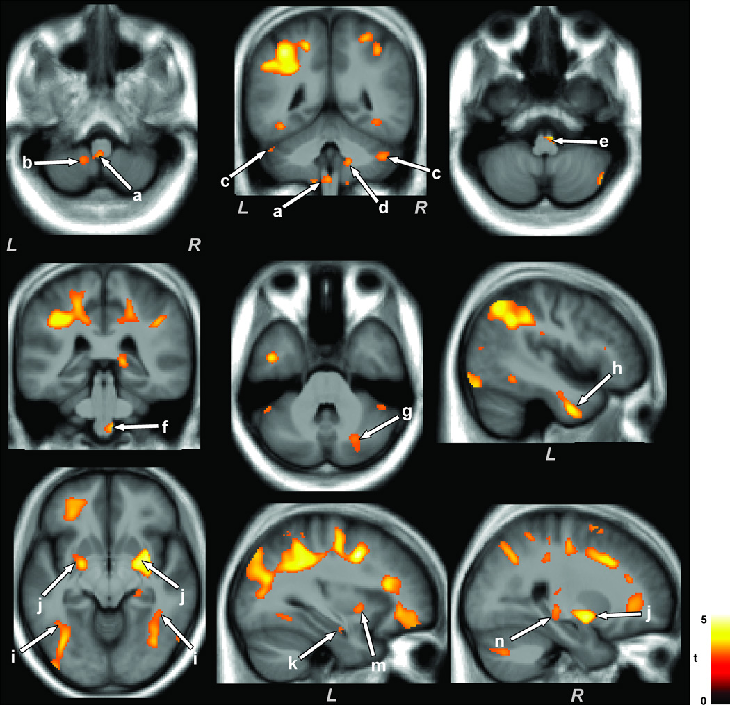
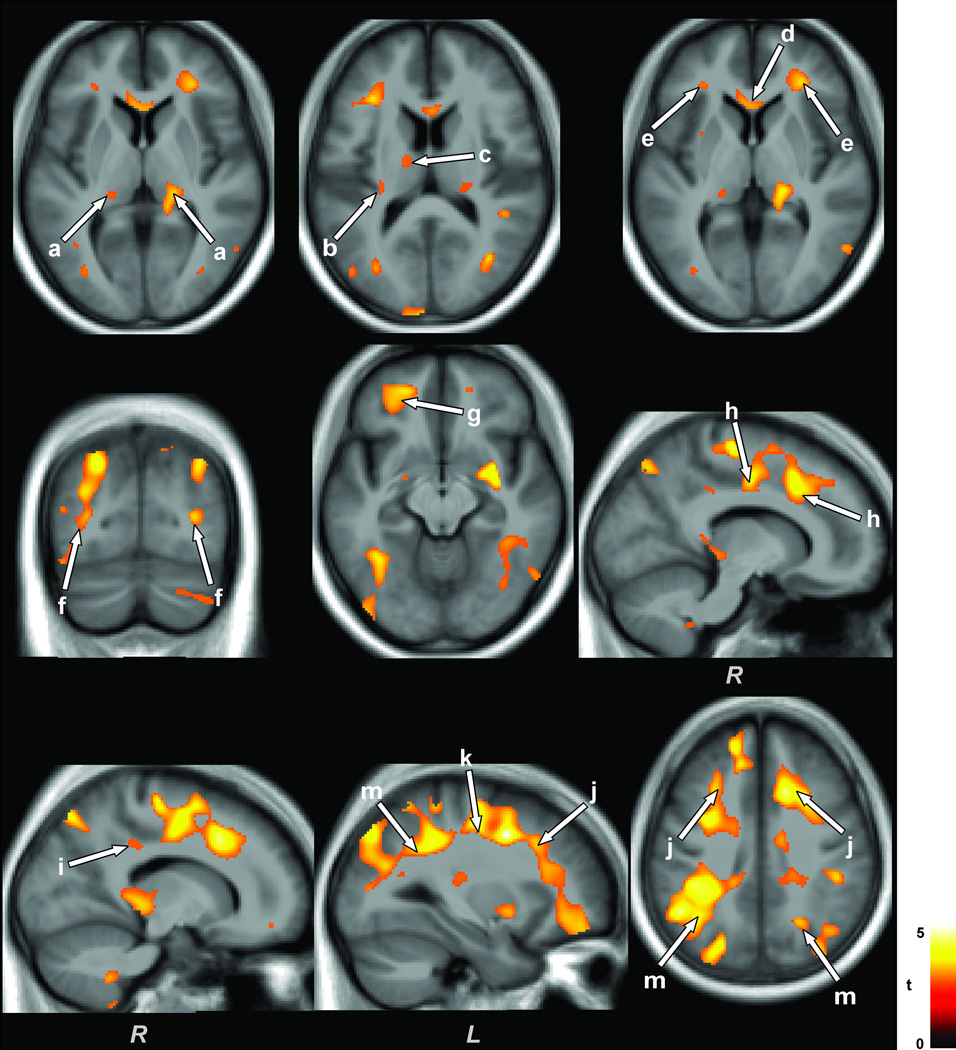
Obstructive sleep apnea (OSA) is a common and progressive disorder accompanied by severe cardiovascular and neuropsychological sequelae, presumably induced by brain injury resulting from the intermittent hypoxia and cardiovascular processes accompanying the syndrome. However, whether the predominant brain tissue pathology is acute or chronic in newly-diagnosed, untreated OSA subjects is unclear; this assessment is essential for revealing pathological processes. Diffusion tensor imaging (DTI)-based mean diffusivity (MD) procedures can detect and differentiate acute from chronic pathology and may be useful to reveal processes in the condition. We collected four DTI series from 23 newly-diagnosed, treatment-naïve OSA and 23 control subjects, using a 3.0-Tesla magnetic resonance imaging scanner. Mean diffusivity maps were calculated from each series, realigned, averaged, normalized to a common space, and smoothed. Global brain MD values for each subject were calculated using normalized MD maps and a global brain mask. Mean global brain MD values and smoothed MD maps were compared between groups by using analysis of covariance (covariate: age). Mean global brain MD values were significantly reduced in OSA compared with controls (P = 0.01). Multiple brain sites in OSA, including medullary, cerebellar, basal ganglia, prefrontal and frontal, limbic, insular, cingulum bundle, external capsule, corpus callosum, temporal, occipital, and corona radiata regions showed reduced regional MD values compared with controls. The results suggest that global brain MD values are significantly reduced in OSA, with certain regional sites especially affected, presumably a consequence of axonal, glial, and other cell changes in those areas. The findings likely represent acute pathological processes in newly-diagnosed OSA subjects.
Copyright © 2012 Wiley Periodicals, Inc.
Conflict of interest statement
Figures
References
-
- Aggleton JP, Vann SD, Saunders RC. Projections from the hippocampal region to the mammillary bodies in macaque monkeys. Eur J Neurosci. 2005;22(10):2519–2530. - PubMed
-
- Ahlhelm F, Schneider G, Backens M, Reith W, Hagen T. Time course of the apparent diffusion coefficient after cerebral infarction. Eur Radiol. 2002;12(9):2322–2329. - PubMed
-
- Ashburner J, Friston KJ. Unified segmentation. NeuroImage. 2005;26(3):839–851. - PubMed
Publication types
MeSH terms
Grants and funding
LinkOut - more resources
Full Text Sources
Medical
