

Leptospirosis in Chonbuk Province of Korea in 1987
- PMID: 2271509
- PMCID: PMC4534998
- DOI: 10.3904/kjim.1990.5.1.34
Leptospirosis in Chonbuk Province of Korea in 1987
Abstract
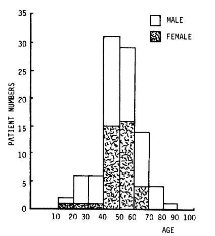
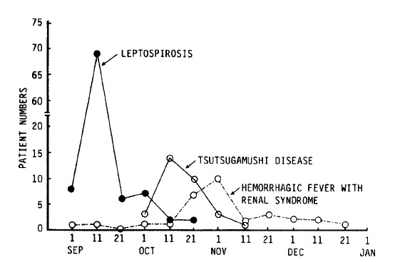
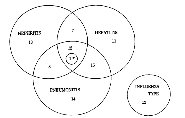
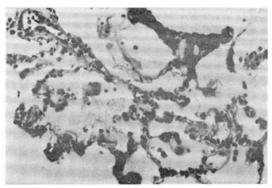
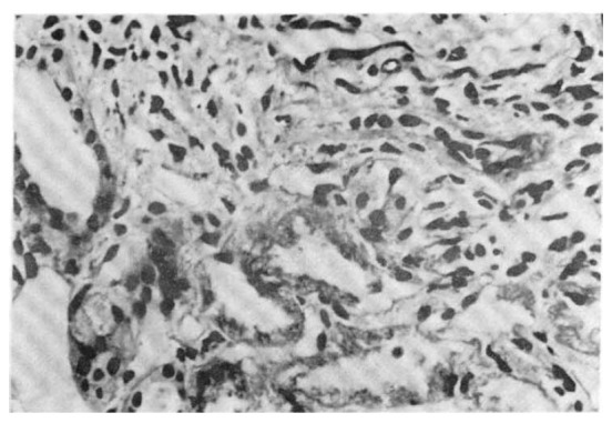
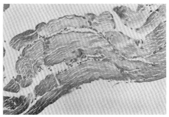
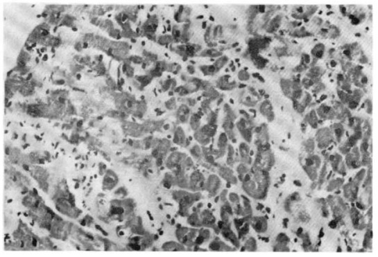
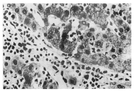
Leptospirosis is a zoonosis with protean clinical manifestations. Its diagnosis requires a high index of suspicion and is confirmed by isolation of the organism or, more commonly, by serologic tests. In the fall of 1987, after severe flooding, we saw 93 patients with leptospirosis, confirmed by a microagglutination test. Thirteen percent of the patients had no clinical or laboratory findings except fever and headache, but the rest had mild to severe manifestations. Jaundice, renal failure, and aseptic meningitis were not common, but pulmonary symptoms, when present, were striking. The mortality rate was 5%. The main cause of death was asphyxiation due to massive hemoptysis from pulmonary hemorrhage and acute respiratory failure.
Figures
Similar articles
-
Leptospirosis in Chonbuk Province of Korea in 1987: a study of 93 patients.Am J Trop Med Hyg. 1989 Sep;41(3):345-51. Am J Trop Med Hyg. 1989. PMID: 2802020
-
Leptospirosis: an ignored cause of acute renal failure in Taiwan.Am J Kidney Dis. 1997 Dec;30(6):840-5. doi: 10.1016/s0272-6386(97)90091-3. Am J Kidney Dis. 1997. PMID: 9398130
-
Pathology and pathophysiology of pulmonary manifestations in leptospirosis.Braz J Infect Dis. 2007 Feb;11(1):142-8. doi: 10.1590/s1413-86702007000100029. Braz J Infect Dis. 2007. PMID: 17625743 Review.
-
Demographic and clinical features of leptospirosis: three-year experience in central Taiwan.J Microbiol Immunol Infect. 2008 Apr;41(2):145-50. J Microbiol Immunol Infect. 2008. PMID: 18473102
-
A retrospective 5-year study in Moldova of acute renal failure due to leptospirosis: 58 cases and a review of the literature.Nephrol Dial Transplant. 2003 Jun;18(6):1128-34. doi: 10.1093/ndt/gfg095. Nephrol Dial Transplant. 2003. PMID: 12748345 Review.
Cited by
-
Clinical Features and Severity of Leptospirosis Cases Reported in the Hawke's Bay Region of New Zealand.J Trop Med. 2021 Jul 6;2021:5567081. doi: 10.1155/2021/5567081. eCollection 2021. J Trop Med. 2021. PMID: 34306102 Free PMC article.
-
Clinical spectrum of pulmonary involvement in leptospirosis in a region of endemicity, with quantification of leptospiral burden.Clin Infect Dis. 2005 Feb 1;40(3):343-51. doi: 10.1086/427110. Epub 2005 Jan 10. Clin Infect Dis. 2005. PMID: 15668855 Free PMC article.
References
-
- Takaki I. The Prevalence of Causative Agents in Rat Bite Fever and Icterohaemorrhagic Spirochetae in Seoul. J Chosun Med Assoc. 1919;28:1.
-
- Ro YM, Yuk SJ, et al. Acute Febrile Hemorrhagic Lung Disease of Unknown Cause. J Korean Med Assoc (in Korea) 1976;19:315–323.
-
- Kim MJ. Clinical Characteristics of Leptospirosis in Korea. J Korean Med Assoc (in Korea) 1988;31:623–627.
-
- Kim JS, Lee JW, Oh Dk, In SD, Lee YH, Cho WH. An Epidemiological Study on Identification of Cause for Epidemic Pulmonary Hemorrhagic Fever. J Korean Med Assoc (in Korea) 1985;28:77–87.
-
- Guidelines for the Control of Leptospirosis. WHO Offset Publ. 1983;67:1. - PubMed
MeSH terms
LinkOut - more resources
Full Text Sources