Comparative Study
. 2012 Aug 17;287(34):28526-36.
doi: 10.1074/jbc.M112.367565. Epub 2012 Jun 19.

Developmental expression of the neuron-specific N-acetylglucosaminyltransferase Vb (GnT-Vb/IX) and identification of its in vivo glycan products in comparison with those of its paralog, GnT-V

Affiliations
Comparative Study

Developmental expression of the neuron-specific N-acetylglucosaminyltransferase Vb (GnT-Vb/IX) and identification of its in vivo glycan products in comparison with those of its paralog, GnT-V

Jin Kyu Lee et al. J Biol Chem. .

Abstract

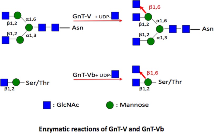
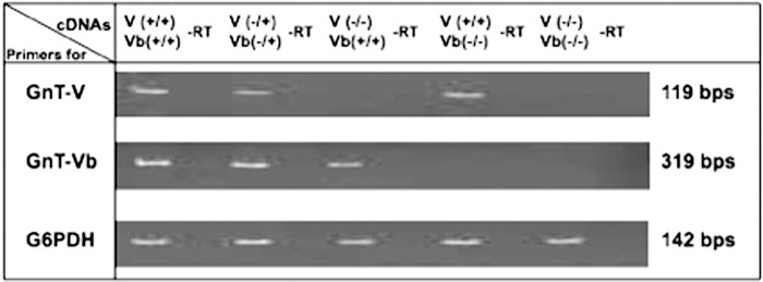
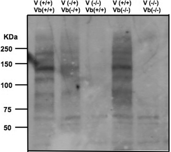
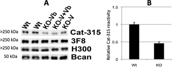
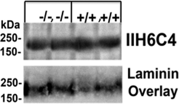
The severe phenotypic effects of altered glycosylation in the congenital muscular dystrophies, including Walker-Warburg syndrome, muscle-eye-brain disease, Fukuyama congenital muscular dystrophy, and congenital muscular dystrophy 1D, are caused by mutations resulting in altered glycans linked to proteins through O-linked mannose. A glycosyltransferase that branches O-Man, N-acetylglucosaminyltransferase Vb (GnT-Vb), is highly expressed in neural tissues. To understand the expression and function of GnT-Vb, we studied its expression during neuromorphogenesis and generated GnT-Vb null mice. A paralog of GnT-Vb, N-acetylglucosaminyltransferase (GnT-V), is expressed in many tissues and brain, synthesizing N-linked, β1,6-branched glycans, but its ability to synthesize O-mannosyl-branched glycans is unknown; conversely, although GnT-Vb can synthesize N-linked glycans in vitro, its contribution to their synthesis in vivo is unknown. Our results showed that deleting both GnT-V and GnT-Vb results in the total loss of both N-linked and O-Man-linked β1,6-branched glycans. GnT-V null brains lacked N-linked, β1,6-glycans but had normal levels of O-Man β1,6-branched structures, showing that GnT-Vb could not compensate for the loss of GnT-V. By contrast, GnT-Vb null brains contained normal levels of N-linked β1,6-glycans but low levels of some O-Man β1,6-branched glycans. Therefore, GnT-V could partially compensate for GnT-Vb activity in vivo. We found no apparent change in α-dystroglycan binding of glycan-specific antibody IIH6C4 or binding to laminin in GnT-Vb null mice. These results demonstrate that GnT-V is involved in synthesizing branched O-mannosyl glycans in brain, but the function of these branched O-mannosyl structures is unresolved using mice that lack these glycosyltransferases.


Figures

SCHEME 1.
SCHEME 1.
Depiction of products of GnT-Vb and GnT-V after typical elongation by additional glycosyltransferases. The GlcNAc residue transferred by each enzyme is highlighted in red.
FIGURE 1.
FIGURE 1.
GnT-Vb is specifically and highly expressed in the developing and adult nervous system. In situ hybridization analyses were conducted on embryos and adult brains to determine the timing and localization of GnT-Vb mRNA. GnT-Vb expression was first detected at embryonic day (E) 7.5 (data not shown), but at all time points it was found almost exclusively within the nervous system. Expression was detected specifically in the nervous system of embryos at E9 (A), E11 (B), and E13 (C). At E15 the expression of GnT-Vb message is shown (D) and the same section was then Nissl-stained (E) to demonstrate the specific localization of GnT-Vb mRNA in neural tissue. F–H, brains from an E15 mouse was removed and sliced horizontally to investigate the spatial distribution of GnT-Vb in the brain. GnT-Vb mRNA (F) and fluorescent Nissl stain (G) of the same section were overlaid (H) to show that GnT-Vb is highly expressed in the subventricular zone (SVZ) and intermediate zone (IZ) and more sparsely in the marginal zone and cortical plate (MZ/CP). It is largely absent from the ventricular zone (VZ). Adult brains were sectioned parasagittally to investigate the spatial distribution of GnT-Vb (I and J) and GnT-V (K). GnT-Vb is highly expressed in the inferior colliculus (IC), nuclei in the thalamus (Thal), the rostral migratory stream (RMS), the dentate gyrus (DG), CA fields of the hippocampus (CA), the striatum and superficial layers of the cortex (I and J). In contrast GnT-V is more broadly expressed throughout the brain although it is largely absent from the striatum (K).
FIGURE 2.
FIGURE 2.
RT-PCR analysis of different genotypes of GnT-V and GnT-Vb K/O mice. Total RNA was isolated from the brain tissues of GnT-V(+/+)Vb(+/+), GnT-V(−/+)Vb(−/+), GnT-V(−/−) Vb(+/+), GnT-V(+/+)Vb(−/−), and GnT-V(−/−)Vb(−/−) mice. Semi-quantitative, real time, one-step reverse transcription (RT)-PCR was performed to amplify a 319-bp GnT-Vb cDNA region containing exons 6 and 7 as described under “Experimental Procedures.” GnT-V expression was analyzed as described and showed a 119-bp PCR product. −RT, no reverse transcriptase reaction. G6PDH (glutaraldehyde-6-phosphate dehydrogenase) PCRs were used as control.
FIGURE 3.
FIGURE 3.
L-PHA-reactive glycoproteins of brain tissue homogenates. Brain tissue samples from adult mice were homogenized in extraction buffer. The proteins were subjected to SDS-PAGE and were blotted to a PVDF membrane. The blot was probed with L-PHA followed by incubation with rabbit IgG against L-PHA and HRP-conjugated goat anti-rabbit IgG.
FIGURE 4.
FIGURE 4.
Western blot analysis reveals that genetic disruption of GnT-Vb reduces the expression of the Cat-315 epitope on RPTPζ. A, postnatal day 0 (P0) brains were analyzed by Western blot analysis with known antibodies against RPTPζ, including 3F8, H300, and Cat-315. H300 detects the RPTPζ protein core, whereas 3F8 and Cat-315 detect specific carbohydrate epitopes on RPTPζ at this age. Brevican (Bcan) was detected to control for protein loading. B, semiquantitative analysis of Cat-315 reactivity from wild types (Wt) and knock-outs (KO). The Cat-315 reactivity relative to brevican has been analyzed as described under “Experimental Procedures.” The combined group of single or double knock-outs were used for KO because they did not differ from each other. n = 4 for wild types and 6 for knock-outs (2 of each genotype).
FIGURE 5.
FIGURE 5.
Analysis of α-dystroglycan glycosylation and laminin overlay assay. Western blot and overlay analysis indicates that double deletion of GnT-V and GnT-Vb does not alter the reactivity of α-dystroglycan to the 116C4 carbohydrate-specific epitope or its ability to bind laminin. Wheat germ agglutinin-enriched samples from wild type (+/+, +/+) or GnT-V and -Vb double knock-out (−/−, −/−) brains were analyzed by Western blot and laminin overlay assays. Each two mice brain tissues were applied. The IIH6C4 antibody detects a specific carbohydrate epitope that is lost from α-dystroglycan in many forms of CMDs and animal models in which O-mannosylation is disrupted. It is, however, unaffected by the absence of GnT-Vb and -Va. The ability of α-dystroglycan to bind laminin seems similarly unaffected.
FIGURE 6.
FIGURE 6.
Nissl-stained brain sections from GnT-Vb and -V double knock-outs show no obvious malformations or changes in cell number or architecture. Thin (3 μm) sections from wild type (Wt) and GnT-Vb and -V double knock-outs were analyzed histologically and using stereological cell counts to determine whether loss of these two enzymes alters brain structure. We found no obvious changes in the morphology of the brain and therefore no changes in the cell number or cellular organization of the brain. These data argue that GnT-Vb and GnT-V are relatively dispensable for normal brain development. Modification of O-mannosyl glycans by these enzymes does not contribute any phenotypes similar to those found in CMDs.

Similar articles

Cited by

References

    1. Dobyns W. B., Kirkpatrick J. B., Hittner H. M., Roberts R. M., Kretzer F. L. (1985) Syndromes with lissencephaly. II. Walker-Warburg and cerebro-oculo-muscular syndromes and a new syndrome with type II lissencephaly. Am. J. Med. Genet. 22, 157–195 - PubMed
    1. Golden J. A. (2001) Cell migration and cerebral cortical development. Neuropathol. Appl. Neurobiol. 27, 22–28 - PubMed
    1. Haltia M., Leivo I., Somer H., Pihko H., Paetau A., Kivelä T., Tarkkanen A., Tom F., Engvall E., Santavuori P. (1997) Muscle-eye-brain disease. A neuropathological study. Ann. Neurol. 41, 173–180 - PubMed
    1. Jimenez-Mallebrera C., Brown S. C., Sewry C. A., Muntoni F. (2005) Congenital muscular dystrophy. Molecular and cellular aspects. Cell. Mol. Life Sci. 62, 809–823 - PMC - PubMed
    1. Lian G., Sheen V. (2006) Cerebral developmental disorders. Curr. Opin. Pediatr. 18, 614–620 - PubMed

Publication types

Substances