Case Reports
. 1990 Jan;5(1):63-8.
doi: 10.3904/kjim.1990.5.1.63.

Pulmonary diffuse alveolar septal amyloidosis--diagnosed by transbronchial lung biopsy

Affiliations
Case Reports

Pulmonary diffuse alveolar septal amyloidosis--diagnosed by transbronchial lung biopsy

C H Kim et al. Korean J Intern Med. 1990 Jan.

Abstract

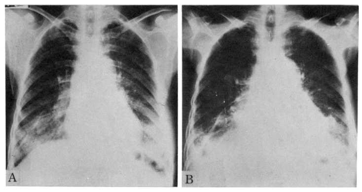
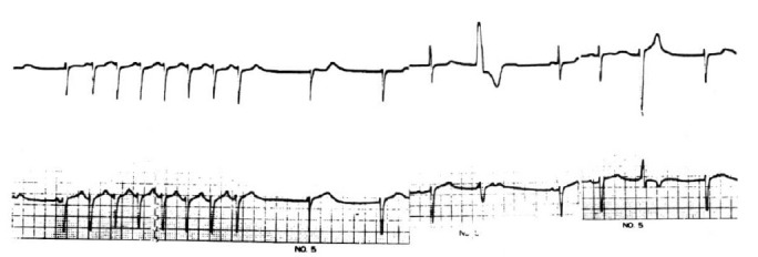
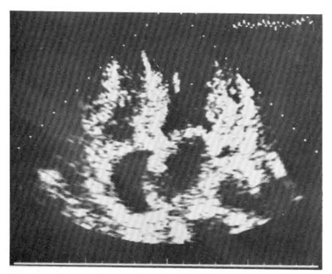
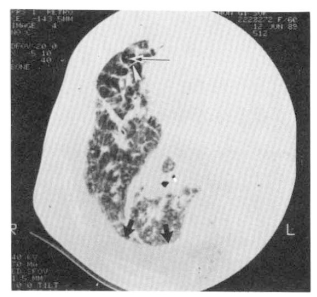
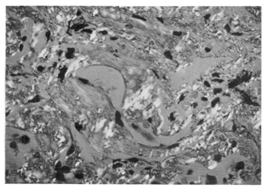
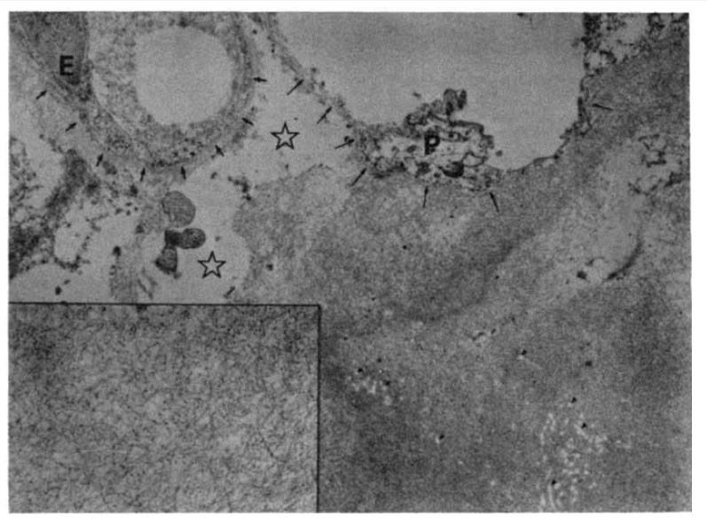
Diffuse alveolar septal involvement is a rare form of pulmonary amyloidosis. Antemortem diagnosis is unusual, and most of the reported cases were diagnosed at autopsy. It has recently been reported that transbronchial lung biopsy via a flexible fiberoptic bronchoscope was a relatively safe method to confirm diffuse alveolar septal amyloidosis. We report a case of pulmonary diffuse alveolar septal amyloidosis confirmed by transbronchial lung biopsy. The patient's chief complaints were dyspnea on exertion and epigastric pain aggravated over a one-year period, while a chest roentgenogram showed bilateral diffuse interstitial infiltration. This case also showed nephrotic syndrome, cardiac arrhythmia, congestive heart failure, a tingling sensation in both hands and multiple nodules in the gastrointestinal tracts, suggesting involvement of the kidney, heart, peripheral nerves and gastrointestinal tracts. We propose that when diffuse interstitial lung disease is present with systemic signs such as nephrotic syndrome or cardiac arrhythmia, amyloidosis should be considered as a possible diagnosis. Also, transbronchial lung biopsy may be a useful confirmative diagnostic tool.


Figures

Fig. 1.
Fig. 1.
Serial chest radiographs of 60-year-old female patient. A. The Initial radiograph shows bilateral diffuse ground glass appearing density, especially in the lower lung fields. B. Radiograph obtained one year later A shows both aggravated lower lung field lesions. Note bilateral pleural effusion and increased heart size as compared with radiograph A.
Fig. 2.
Fig. 2.
A Holter ECG recording shows sinus bradyarrhythmia with junctional escape rhythm, episode of paroxysmal supraventricular tachycardia (PSVT) and multiform ventricular premature beats (VPBs).
Fig. 3.
Fig. 3.
A two-dimensional echocardiogram shows a thickened left ventricular posterior wall, interatrial and interventricular septum. The myocardium shows an increased refractile pattern, a so-called “granular sparkling” appearance.
Fig. 4.
Fig. 4.
A high resolution CT obtained at right lower lung field (retrospective retargeting, FOV 20 cm, 140KVP, 510 mAs, 512×512 matrix, bone algorithm) shows prominent interlobular septa (white arrowhead) and a central core structure (thin arrow), as well as a diffuse, ill-defined, increased hazy density. Increased density between the rib and the lung (thick black arrows) is due to pleural effusion.
Fig. 5.
Fig. 5.
Polarizing microscopy following Congo-red stain reveals apple-green birefringence at the vascular walls and along the alveolar septa (×400).
Fig. 6.
Fig. 6.
Electron microscopic observation of the lung reveals massive deposition of fibrillar material in the alveolar septum. The alveolar basement membrane (long arrows) is incorporated into the amyloid substance. Retractile white spots and fibers (tiny arrowheads) in the amyloid pool are preexisting collagen fibers. Free space (stars) in the interstitium is due to interstitial edema. Alveolar epithelial cells (P) are artifactually deformed. (E: endothelial cells; smaller arrows: capillary basement membrane). (Bar represents 1 um, ×14,000) (Inset) Magnification of the amyloid substance reveals nonbranching fibrills of approximately 100 nm in length and 10 nm in diameter. Granular deposits, less than 10 nm in diameter, are scattered also. (Bar represents 0.1 um, ×56,000).

References

    1. Glenner GG. Amyloid deposits and amyloidosis: The beta fibrillosis. N Engl J Med. 1980;302:1283–1292. 1333–1343. - PubMed
    1. Cohen AS, Shirahama T, Sipe JD, Skinner M. Amyloid proteins, precursors, mediator, and enhancer. Lab Invest. 1983;48:1–4. - PubMed
    1. Kyle RA, Greipp PR. Amyloidosis (AL): Clinical and laboratory features in 229 cases. Mayo Clin Proc. 1983;58:665–683. - PubMed
    1. Kanada DJ, Sharma OP. Long-term survival with diffuse interstitial pulmonary amyloidosis. Am J Med. 1979;67:879–882. - PubMed
    1. Poh SC, Tjia TS, Seah HC. Primary diffuse alveolar septal amyloidosis. Thorax. 1975;30:186–191. - PMC - PubMed

Publication types